There are different types of moles, but not all of them are comfortable or safe. Birthmarks are convex and round, may have a black or yellowish tint, and hair may grow on them. In addition to being an aesthetic drawback, moles are prone to proliferation and are considered a harbinger of skin cancer. Therefore, it is recommended to remove them. There are various methods for removing moles, each of which has its own characteristics, advantages and disadvantages.
There are various methods for removing moles. The most effective method is selected by the doctor individually, taking into account the number, location, nature and size of skin tumors.
In what cases is the removal of moles indicated and what method of destruction of skin tumors is better to choose? We’ll talk in the article.
Indications and limitations for mole removal
Surgical removal of a nevus is prescribed if there are the following indications:
- Presence or suspicion of cancer;
- Nevi that have broken into several parts;
- Moles of large area or size.
Surgical removal of a mole, like any surgical procedure, has a number of limitations. These include:
- Herpetic infection in the active stage;
- The presence of inflammatory diseases;
- Infectious diseases.
Removal of nevi can be carried out after elimination of the clinical manifestations and causes of the listed pathological processes, that is, after the patient has recovered.
It is strictly forbidden to remove nevi on your own, as this can cause infection of the body and the development of oncology. Surgical excision of a mole should only be performed in a clinical setting by a qualified specialist.
Is the appearance of moles a natural phenomenon?
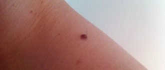
A mole is a small pigmented area of skin that indicates congenital defects or acquired cancer. The size of such formations can vary from several millimeters to tens of centimeters. Moles can also have different pigmentation - from slightly reddish to black. They may or may not be convex, they may or may not have hair growing on them - in any case, any such manifestation is normal from a medical point of view.
Important ! The only sign that it’s time to worry is the appearance of moles after a long exposure to the sun or when they appear and disappear regularly. If this occurs, you should immediately consult a doctor.
A mole is most often a normal phenomenon
Advantages and disadvantages of surgical removal of nevi
Despite the fact that surgical removal of nevi is the oldest method among all known, it has a number of undeniable advantages over others:
- Possibility of getting rid of deep and large moles;
- Safe excision of malignant neoplasms;
- The procedure is painless, since all manipulations are performed using anesthesia;
- Affordable price;
- One hundred percent removal of the nevus in one session (after excision, no neoplasm cells remain, which eliminates the reappearance of the mole);
- Possibility of conducting histological examination of remote formation;
- No contraindications.
Excision of a mole using the classical method is an effective and safe way to get rid of a nevus. However, despite this, it has several disadvantages:
- High likelihood of keloids and scars;
- Long period of rehabilitation;
- After surgery, you should not be in the sun.
Traditional removal methods
The Internet is full of recipes and various methods that are considered effective in removing moles and papillomas. However, their benefits remain controversial - using them risks only aggravating the situation. That is why you should not trust the dubious advice of a complete stranger who does not provide any confirmation of his words besides personal experience.
It's better not to use folk remedies
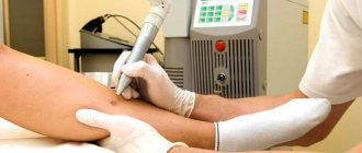
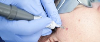
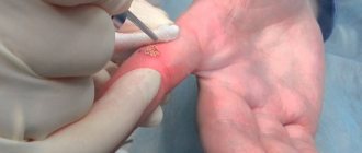
How is surgical removal of moles performed?
There are two methods for surgical removal of a mole: excision with cauterization (burning out using a special device) and excision followed by suturing. The operation lasts from 40 to 60 minutes. The duration of the manipulation will depend on the individual characteristics and condition of the patient, as well as the size and location of the mole.
Excision of nevi without cosmetic sutures consists of the following steps:
- Treatment of the operated area with antiseptic drugs;
- Carrying out local anesthesia;
- Excision of the nevus using a scalpel at the skin level;
- Cauterizing the wound with a special device for cauterization or applying a drug for local use;
- Treatment of the wound with a local antibacterial agent;
- Applying a sterile dressing.
Upon completion of the operation, the doctor provides comprehensive recommendations for caring for the postoperative area and sends the resulting material for histological examination.
Excision of a mole with suturing is carried out in several stages:
- Treatment of the operated area with antiseptics;
- Use of local anesthesia;
- Excision of the nevus with the capture of surrounding healthy tissue;
- Treatment of the wound with antiseptic drugs;
- Cosmetic stitches. Depending on the depth of the formation, superficial, self-priming and deep sutures can be used;
- Treatment of the wound with a local antibiotic;
- Applying a patch and sterile dressing.
The removed material is also sent for histology to confirm the benignity of the neoplasm. If the nevus turns out to be malignant, then another intervention is performed. A week after the operation, the sutures are removed (if superficial sutures are used).
Recently, surgeons have been using special silicone absorbable patches, which make the scar left after excision less noticeable. They are glued to the scar and worn for 15-20 days. In addition, if the patient is not satisfied with his appearance, plastic correction can be performed in the future.
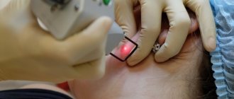
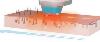
Laser removal of hemangioma
Today, the best method for removing hemangiomas is laser removal. During removal, a laser beam is applied to the tumor and it evaporates. Laser removal is bloodless, since the beam coagulates all vessels. During removal there is no contact between the instrument and the wound, which eliminates wound infection. Removal of hemangiomas using a laser can be performed on an outpatient basis under local anesthesia. During removal there is no pain at all. On average, the removal procedure lasts from 2 to 15 minutes; for larger sizes, several procedures may be required.
Make an appointment
Rehabilitation period after mole removal
Surgical excision of nevi is associated with the longest period of rehabilitation. Depending on the structure, location and size of the nevus, the recovery period can last several weeks.
Throughout the rehabilitation period, the patient should adhere to the following recommendations:
- Do not scratch or tear off the crust that covers the postoperative wound;
- Do not sunbathe on the beach or in the solarium;
- Avoid getting water on the wound for a week after the intervention;
- When excising plantar nevi, do not subject the operated area to excessive stress. This will prevent possible bleeding;
- Carry out independent replacement of postoperative dressings only with the permission of the doctor;
- Follow hygienic skin care rules.
Also, rehabilitation after excision of a nevus involves cosmetic surgery to eliminate visible scars.
Reviews
Irina, Saratov
“I was removing a mole, which did not affect my health in any way, it was simply in the way because it was constantly clinging to the chain hanging on my neck. I decided to remove it with liquid nitrogen - it was quick, the rehabilitation was short, and the price was reasonable. I came, having previously visited all the necessary doctors. They treated me, burned me, and told me not to go out in the sun, but this was understandable - it was late winter outside. The wound healed faster than in 2 weeks - after all, the mole was small. A small light spot remains, but it is hardly noticeable on the skin. I am very pleased with the result, I recommend it to anyone who asks for advice. Six months have passed, and I have never regretted it.”
People are happy with the procedure
Ksenia, Voronezh
“I had a mole removed surgically because... There were suspicions of cancer. Fortunately, they were not confirmed. I didn't like that mole anyway, so I'm glad it's gone. I understand that this is a rather “outdated” method, but there was a chance to send the material for examination. There is a small scar left, but it seems only I can see it. The doctor did everything very carefully, there were no complications. So I advise you not to worry and, if necessary, remove such tumors.”
Nikolay Kolyuzhny, surgeon
“Never treat choosing a clinic with disdain, do not leave everything to the last minute. It is very important that the doctor is an expert in his field, because removing a mole is a process that requires certain knowledge and specific skills. The wrong attempts to solve a problem can backfire in the future and lead to problems that will be incredibly difficult to fix.”
The fourth scenario is casuistic
It was removed, the histology was good, but skin cancer (melanoma) developed at the site of removal.
A benign mole was removed; according to histology, it was a nevus; the histology was reviewed in three laboratories, but a non-pigmented melanoma developed at the site of removal, which was also reviewed several times in several institutions. A very experienced colleague told me this story. How can this even be? I think the reason is the following.
As we know, melanoma is a tumor that develops from melanocytes. These cells are evenly distributed throughout the skin. Thus, a tumor can develop on almost any part of the body, and the site of mole removal is no exception. Most likely, here we are also dealing with a coincidence, the extremely low frequency of occurrence of which only proves its probability.
How to prevent this scenario? It’s very simple: avoid risk factors for melanoma.
Photos before and after
Photos before and after laser mole removal
Photos before and after mole removal with liquid nitrogen
Photos before and after surgical removal of a mole
Photos before and after radio wave excision of a mole
Photos before and after electrocoagulation of a mole
ABCD dermoscopic rule
In dermatology, there is the ABCD rule, which represents diagnostic signs that allow one to clearly limit a particular structure of a mole.
Read also Cutaneous leishmaniasis - symptoms, diagnosis, treatment
A – asymmetry. Benign formations have a symmetrical shape, that is, one half is similar to the other.
B – side, border. Benign formations have clear, even boundaries, without any irregularities, outgrowths, or processes.
C – color, color. The color of the mole should be uniform. If there are any inclusions, the appearance of red or blue shades, then you need to think that something wrong is happening to the mole.
D – formation diameter. If the diameter increases throughout life, it increases evenly.
In what cases should a mole not be removed?
The first and most important rule should be that independent removal of any moles is strictly prohibited. Damaging the surface of the formation can provoke the development of a cancerous tumor. Based on a thorough examination, your doctor may or may not recommend laser mole removal surgery. A conclusion with a ban is made if the nevus is oversized, located in an inconvenient place and there is no guarantee that it will be completely removed. Otherwise, there is a certain risk of getting blood poisoning or further growth of the formation.
There are a number of diseases for which laser mole removal is contraindicated:
- diabetes;
- pregnancy in all trimesters;
- oncological diseases;
- mental and nervous disorders;
- herpes rash in the immediate vicinity of the nevus;
- HIV.