It is possible to look fresh at any age! How? The answer is eyelid canthopexy. The factors that most often contribute to this are aging of the body and ultraviolet radiation. Youth, alas, is not eternal. Although there is hardly a person who would not want to detain her. The elasticity of the skin decreases with age, causing the lower eyelids to droop, resulting in bags under the eyes. However, it is not uncommon for bags to appear at an early age. As a result, we have a fading effect on a relatively young face.
Medical indications
In addition to aesthetics, there are also medical indications, such as:
- Myopia (and as a result, bulging eyes);
- Consequences of chronic inflammatory processes;
- Weak muscles that do not hold the corner of the eye;
- Ectropion (inverted lower eyelid)
- Ankyloblepharon (partial fusion of the eyelid);
- Incomplete cleavage;
- Blepharophimosis (narrowed incision);
- Chronic problems with the conjunctiva.
All of the above is a reason for an immediate trip to the surgeons.
Preparing for surgery
It is very important to consult with an ophthalmologist, since this area of influence is his diocese. Afterwards it is necessary to undergo a series of tests in order to identify contraindications. This:
It is necessary to be examined at the preparation stage in order to identify contraindications to the procedure. But everyone who wants to undergo the procedure must know about the diseases due to which canthoplasty is not permissible:
- blood diseases (clotting disorders) impaired clotting;
- pathologies of the endocrine system;
- high pressure inside the eye;
- “dry” eyes;
- severe myopia;
- oncology;
- pregnancy and feeding.
To compare the results, surgeons take photographs of the face in general and specifically the eyes from different angles. There are specialized computer programs for preliminary visualization of the results of the upcoming intervention.
How are the operations performed?
Before both operations, the patient must consult with the surgeon and ophthalmologist. You may have to undergo general and biochemical blood tests to rule out the presence of inflammation in the body. The appropriate anesthesia method is also selected.
When choosing canthopexy, the intervention proceeds through the following stages:
- the patient is given local anesthesia or general anesthesia, and the eyeball is protected with a plastic shield; incisions are made in the natural folds (preferably): 1-2 mm below the eyelash line of the lower eyelid and 0.7-1 cm under the eyebrow, in the area of the outer third ;
- Through a subglacial incision, the edge of the periorbital muscle is sutured and tightened with a self-absorbing thread. The seam ends up inside;
- Excess skin is cut off and a suture is applied.
In canthoplasty, the intervention follows a similar scheme, but with one difference: a small fragment of the lateral tendons is resected, the ends of the tendons are sutured and fixed to the periosteum.
On average, each operation lasts about one and a half hours. The time increases if additional interventions are carried out in parallel. As a rule, the patient goes home a few hours after the operation.
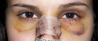
The recovery period lasts 2-3 weeks. On days 5-7, the external suture is removed. Before the suture is removed, you need to take analgesics prescribed by your doctor every 7-8 hours. Throughout the entire rehabilitation period, swelling, hematomas, irritation of the conjunctiva, tearing, and hardness in the corners of the eyes may be present. For 1-2 weeks, it is possible to use softening and moisturizing eye drops.
Before the end of the rehabilitation period, you should reduce eye strain: work less on the computer, watch TV, read. You should also avoid exposure to the sun, visiting baths and saunas, and do not use contact lenses or cosmetics. Do not bend over or lift heavy objects. It is recommended to sleep on your back, placing a high, flat pillow under your head.
Obstacles to canthoplasty or canthopexy may include:
- dry eye syndrome;
- diabetes mellitus and other endocrine disorders;
- pregnancy and breastfeeding;
- cardiovascular diseases;
- cancer diseases;
- inflammation of the eyes, eyelids;
- glaucoma;
- high blood pressure.
Possible complications after surgery are wound suppuration, bleeding, and the appearance of scars. However, such cases are quite rare.
Technique of canthopexy surgery
The duration of the surgical intervention is up to two hours. The canthus that supports the corner of the eyelid moves and is fixed in a new place, as a result the eye opens and rises. The lower eyelid is adjacent and in a normal position.
The operating room is always sterile. An incision up to 1 cm long is made inside the natural crease of the eyelid and is completely invisible. Next, the canthus is separated from the bone, the tendon is stretched and fixed, excess tissue is excised if necessary, and then the incisions are sutured with very thin threads.
Local anesthesia. You can go home immediately after the operation. In case of pain, the medical staff gives an analgesic. But, despite the fact that canthopexy is considered a minor operation, you still need to go to the clinic not alone, as you may need the help of loved ones after it.
Most popular: Does your facial skin need thermal water from the depths of the earth?
Side effects include pain, bruising, and swelling due to damaged nerves and damaged blood vessels. It is also possible for infection to get into the seam.
Rehabilitation
Complete rehabilitation after surgery takes up to one month. As for the active recovery period, it lasts about a week.
During this time, the bruises and swelling that are inevitable when intervening in the periorbital area have time to completely disappear. The eyes regain their normal appearance. You can safely go to work.
Within 7-10 days, the following recommendations must be strictly followed:
- Until the stitches are completely healed, treat them with an antiseptic.
- Sleep only on your back with your head slightly elevated.
- Do not wear contact lenses.
- Do not use decorative cosmetics.
- Do not overstrain your eyesight, work less at the computer.
- Avoid significant physical activity.
- Quit smoking and alcohol, minimize coffee consumption.
- Do not visit the sauna and solarium, do not overheat in the bathroom.
- Protect your eyes from the sun with dark glasses.
Ignoring doctors' recommendations can lead to problems. The most common among them are suture dehiscence and the appearance of post-traumatic pigmentation.
Causes and origins of drooping eyelids
Our age and problems are revealed precisely by the “mirrors of the soul.” The skin around them is softer and thinner, the muscles are weak, the ligaments are more fragile. That's why even older people experience crow's feet. In general, this is a sign that a person smiles often. But optimists are also reluctant to grow old prematurely. But over the years, this “stamp of fun” turns into drooping eyelids, which, on the contrary, give the face a sad and tired expression, like a crooked nose.
The canthus has long thin tendons on the outside, which weaken and stretch over time, resulting in the corners of the eyes creeping down. Sometimes people are born with such eyes - if the tone of the tendons is very weak by nature.
Reasons from an anatomical point of view
The periorbital (circular) muscle, located under the skin around the eyeball, closes the eyelids. When it contracts, the outer corner wrinkles like a fan. The constant load on the muscles around the eyes overstretches the skin, which is why wrinkles remain even at rest. And with age, their number increases, as skin turgor decreases.
The soft cartilage under the orbicularis muscle is held in place by connective tissue and tendons. With one edge they are connected to each other, with the other they are attached to the side of the cartilage. This forms the angle of the eye - the canthus, fixed to the periosteum. In young people, the corners of the eyes are at the same level or the outer corner is slightly higher than the inner. With age, the outer edge droops.
The most convex places of the eyeballs and the highest points of the cheekbones for many people are at the same level and form a line called the “neutral vector”. The displacement of the protruding part of the cheekbone forward relative to the point of the cornea is a “positive vector”. They are also called normophthalmos - these are signs of the proper development of bones and tissues supporting the eyelid.
If underdevelopment of the facial skeleton or tissue failure is observed, the vector becomes negative - the eye protrudes beyond the orbit (bulging eyes, also known as exophthalmos). The eyeball can become this way due to certain diseases (thyrotoxicosis, Graves' disease, brain tumor). But more often it’s just a sign of age.
Gravity affects everything – including the skin of the eyelids. Sagging cheeks and lower eyelids are called ptosis. If the eyelid loses support, it recedes downwards from the eye. The result is a displacement of the outer angle to a level lower than the inner one, causing the mechanical support of the eyeball to be partially lost.
The eyes widen, the lower eyelid turns forward, and the sclera becomes visible. If this phenomenon progresses, the mucous membrane of the eye falls out. In this case, conventional blepharoplasty is powerless. And then external canthopexy comes to the rescue. In addition to the above effects, canthopexy also eliminates facial asymmetry and other pathological changes and aesthetic visual defects.
Most popular: Does your facial skin need thermal water from the depths of the earth?
What is the difference between canthopexy and canthoplasty?
The terms “canthopexy” and “canthoplasty” are often used interchangeably, but this is incorrect. They represent different, albeit similar, interventions.
Canthoplasty is an operation to correct the shape of the eyes. During the intervention:
- through the periorbital muscle, the tendons supporting the external canthus are separated from the bone;
- part of the tendons is removed or a ligature is used, which allows you to correct and fix the muscles and ligaments without resection;
- they tighten the inner part of the lower eyelid and attach it to the place where the tendons used to be;
- remove excess eyelid skin.
Canthoplasty can be lateral or medial. In the first case, ptosis of the outer corner of the eye is corrected, in the second - the inner corner of the eye.
This procedure is often used not only to correct age-related changes, but also to give the eyes a European or Asian shape.
Unlike canthoplasty, with canthopexy the surgeon tightens the outer canthus without partial removal or relocation, but only by suturing the tendons.
In addition, canthopexy often becomes an additional procedure in transconjunctival or laser blepharoplasty, for example, after removal of fatty hernias. The procedure can also be combined with a circular facelift, lipolifting and other interventions.
Thus, we can say that canthoplasty is a more complex type of canthopexy. That is why the first procedure is more often used to correct medical defects, and the second - aesthetic ones. The effect of both operations lasts for 5-10 years, depending on the age and condition of the patient’s skin.
Canthopexy and canthoplasty have the following goals:
- eliminate sagging of the lower or upper eyelid;
- correct eversion of the eyelid (ectropion) and excessively round eyes (this effect can be observed with endocrine disorders);
- prevent the “bulging eyes” symptom in people with myopia;
- correct facial asymmetry with facial paralysis;
- correct blepharophimosis - shortening of the eye shape;
- remove signs of aging.
Difference from canthoplasty
Canthoplasty and canthopexy are elements of blepharoplasty. But there are differences between them. Canthopexy – the canthus is tightened.
It is believed that there is no fundamental difference between these two surgical procedures. We can say that one of them is a variety of the other, and vice versa. The principle is the same - surgical action on soft tissue and cartilage. With the help of these procedures, you can make eyes Asian, or vice versa – European, at the request of the patient. In addition to removing excess skin, hernial elements from the infraorbital bags are also eliminated. The lateral area is affected when it is fixed to the periosteum. The sutures are made with a non-absorbable thread, the ends of which are tied and tightened. The result is two barely noticeable nodules on the skin. Canthoplasty – the excess is partially removed and stitched back together.
Indications
Canthopexy is required in the following cases:
- Drooping of the corners of the eyes with age;
- Genetically determined drooping eyelid;
- Flabby and stretched eyelids;
- The patient’s desire to achieve a “cat-like look”;
- Bulging eyes;
- Eversion of the eyelid as a result of paralysis;
- during the process of blepharoplasty.
Some diseases not related to aesthetics can also be treated with canthopexy.
Contraindications
This procedure is not possible if:
- inflammation of the eyes (conjunctivitis);
- poor blood clotting;
- chronics in the acute stage;
- diabetes mellitus during the period of decompensation.
Any of the noted diseases does not make it possible to canthopexy the eyelids.


