Causes of serous mastitis
In the serous form of mastitis, the parenchyma and milk ducts of the gland become inflamed. The infiltrate does not consist of pus, but of lymph and intercellular fluid.
Causes of the disease:
Incorrect hygienic breast care during breastfeeding - rubbing the nipple, infrequent or too frequent wiping or washing of the breast.
Installation of an artificial implant, compression with synthetic material, disruption of lymph outflow.
Incorrect grip of the nipple during feeding: the baby pulls it without covering part of the areola with his mouth.
Stagnation of milk and disruption of its normal outflow.
Stagnation of milk, or lactostasis, leads to impaired absorption and secretion of lactic acids. As soon as the serous fluid penetrates the breast tissue, the outflow of lymph becomes difficult and an infiltrate forms. The appearance of a serous infiltrate leads to inflammation of the gland tissue. If infectious microorganisms penetrate into the breast through damage in the form of cracked nipples, serous mastitis transitions into a purulent form.
Complications of serous mastitis
The entry of pathogenic bacteria into the infiltrate causes an adequate response of the immune system - the formation of pus.
Complications:
- Formation of an abscess - an encapsulated collection of pus;
- The formation of phlegmon - diffuse inflammation of the glandular tissue of the breast,
- Dilation of the milk ducts, which subsequently prevents normal lactation and threatens suppuration.
Diagnosis of serous mastitis
In order not to give up breastfeeding, you need to detect signs of the disease in time. The doctor should know exactly when breast problems began. The doctor should monitor changes in body temperature. If it is elevated, general antibacterial therapy is carried out using broad-spectrum antibiotics.
On palpation, a painful lump on the chest is noted. In some cases, clear liquid is released from the nipple even with slight pressure on it.
Another diagnostic method is ultrasound. With this research method, it is possible to accurately determine the location of the infiltrate or the presence of lactostasis.
Pleural fluid accumulation syndrome due to inflammation
The accumulation of fluid in the pleural cavity can be triggered by an inflammatory process. In this case, doctors talk about exudation (exudation of effusion in the form of exudate). The mechanism of this pathology is caused by an infectious lesion and includes the following changes in the body:
the permeability of the vascular walls is increased,- blood overflow of tissues in the area of inflammation,
- increased oncotic pressure,
- symptoms of a primary inflammatory disease make themselves felt.
The pleural cavity can be filled with the following types of inflammatory effusion:
- Serous. Transparent liquid. It is released during inflammation of the serous layers of the pleura. The prognosis is favorable. Sources of inflammation are burns, allergies, viruses. For example, pleurisy is accompanied by effusion of serous exudate.
- Fibrous. More dense, villous exudate, with an increased content of fibrin. The pleural membrane is destroyed under the influence of this fluid: scars, adhesions, and ulcers appear.
May be released due to tuberculosis. - Purulent. Opaque, viscous liquid in the pleural cavity of a green hue. Consists of a large number of spent protective leukocyte cells. Caused by the entry into the body of pathogens such as fungi, streptococci, staphylococci.
- Hemorrhagic. It occurs as a consequence of the destruction of blood vessels. It is a reddish liquid due to its saturation with red blood cells. Occurs in tuberculous pleurisy.
Treatment emphasizes antibacterial drug therapy and is aimed at destroying the infectious agent. To remove exudate, they resort to surgery.
Mastitis in a nursing mother
In relation to the total number of births, the frequency of mastitis ranges from 3 to 20%. In most cases, purulent inflammation in the mammary gland in women in the postpartum period is caused by S. aureus (Staphylococcus aureus). The “entry gates” for pathogenic microbes are cracks and microtraumas of the nipples. It is possible that infection can enter through the milk ducts during feeding or pumping. Mastitis in a nursing mother can be a consequence of improper care of the mammary glands or develop due to non-compliance with personal hygiene rules.
Postpartum mastitis, unlike its other forms, is predominantly associated with lactation (hence the name “lactation”) and is diagnosed in 2-11% of lactating women. Lactation mastitis is characterized by unilateral damage to the mammary glands, develops mainly at 5-6 weeks after birth and goes through all the stages inherent in acute purulent mastitis of any origin.
Violation of the regimen and/or rules of breastfeeding provokes stagnation of milk in the mammary gland, which provokes the development of local non-infectious inflammation.
Since the trigger for the development of inflammation is lactostasis, at the beginning of the disease a woman experiences a feeling of tension in the mammary gland. Stagnation of milk leads to the fact that the mammary gland increases in size, and overcrowded milk ducts can be felt in the form of painful compactions without clear boundaries. The amount of expressed milk decreases significantly and body temperature rises.
If lactostasis is not eliminated in the next 3 to 4 days, secondary pathogenic flora attaches, which causes decomposition of milk and damage to the milk ducts, that is, the process takes on the character of acute purulent inflammation. The mammary gland looks swollen and red, the discharge from the nipple becomes purulent, and symptoms of intoxication increase. An attempt to empty the mammary gland is not possible due to severe pain. The further scenario of the disease depends on how quickly the patient seeks qualified help: if a woman does not turn to a specialist, does it too late, or tries to cope with the disease on her own, the likelihood of developing a severe infectious process becomes very high.
Non-lactation mastitis after childbirth is much less common; it develops without the participation of lactation and is similar to that in non-lactating women.
Manifestations and key principles of disease therapy
Serous mastitis is only one of the first stages of the disease, which without timely treatment can turn into an infiltrative and later purulent form.
Symptoms of mastitis occur quickly, within just a few hours. The main symptoms of serous mastitis include:
- a sharp increase in temperature to 38-39.5 °C;
- general weakness, chills, feeling “broken”;
- breast enlargement, heaviness, chest pain;
- redness and infiltration of the breast;
- headaches, nausea, vomiting.
Increased risk factors for serous mastitis are:
- large breasts;
- cracked and irregularly shaped nipples;
- chronic inflammatory diseases;
- decreased immunity;
- the occurrence of lactostasis.
Lactostasis is stagnation of milk in the ducts due to the appearance of “plugs” from thickened secretory fluid. With lactostasis, small lumps appear in the breasts, which disappear after massaging. The breasts begin to hurt, and milk production becomes uneven. If lactostasis is not cured within a few days, it turns into mastitis.
Serous mastitis is usually easily diagnosed. The doctor examines the mammary glands and asks clarifying questions regarding symptoms and the timing of their onset. Then additional studies are prescribed, which may include:
- general blood test to identify the inflammatory process;
- examination of breast milk to determine the type of pathogen and response to antibiotics;
- Ultrasound, computed mammography or thermography of the mammary glands to exclude other diseases, for example, mastopathy or oncology.
Treatment of serous mastitis is conservative. It is based on antibacterial therapy with drugs (Cefazolin, Cefoxitin, Ceftriaxone). In addition, a whole range of auxiliary measures is used:
- restriction of physical activity and exercise, adherence to bed rest to prevent cardiovascular problems, which can manifest themselves as a complication of serous mastitis;
- refusal to wear a compressive bra, which disrupts the outflow and can cause stagnation of milk;
- regular expression of milk to prevent stagnation, bacterial growth and compression of adjacent ducts and vessels of the mammary gland;
- partial suppression of milk production by reducing the amount of fluid consumed or using special medications prescribed by a doctor;
- physiotherapy on the second day after suppression of acute symptoms: microwave therapy, ultrasound, ultraviolet irradiation.
Classification of mastitis
Mastitis is classified as follows:
With the flow:
Acute - characterized by all the clinical manifestations of mastitis, with severe chest pain.
Chronic – characterized by a weakening of the clinical manifestations of the disease with periodic exacerbations. Upon palpation, a dense, inactive seal fused to the skin is determined.
Forms of mastitis
Lactation mastitis (postpartum) is primarily a consequence of prolonged lactostasis during breastfeeding. It is characterized by breast swelling, sharp pain when feeding or expressing milk, redness of the skin and a rise in body temperature. The milk may contain impurities of pus and blood.
Plasma cell mastitis is a rare type of disease caused by multiple births. It develops mainly after lactation and in women after 30-40 years. It is characterized by processes of infiltration of tissues under the nipple with plasma cells, and is also accompanied by processes of hyperplasia of the excretory mammary ducts. There is usually no formation of pus. Clinical manifestations may resemble breast cancer.
Mastitis of newborns - just like in adults, it is characterized by swelling of the mammary glands with discharge when pressed, only not only in girls, but also in boys. Inflammation is caused by the residual effect of sex hormones transmitted from the mother. Clinical manifestations usually subside after a few days, however, in some cases medical attention may be required.
By the nature of inflammation
- Serous is the initial stage of the development of pathology, during which serous fluid appears in the mammary gland.
- Infiltrative is the next stage of the development of the disease, in which serous fluid from the mammary gland seeps into the external tissues, forming foci of infiltration, while the breasts become full and the skin becomes stretched.
- Purulent is the third stage of mastitis development, in which infiltrates are transformed into purulent deposits.
- Abscess – characterized by a limited purulent focus in one place, localized.
- Phlegmonous - characterized by widespread purulent formation in the chest.
- Gangrenous – characterized by the formation of necrotic foci during a long course of a purulent inflammatory process.
Accumulation of edematous fluid in the pleural cavity
The fluid between the pleural layers can increase in volume regardless of inflammatory processes. In this case, its accumulation is due to a failure of the natural process of its production or reabsorption.
For such cases, the term “transudate” (non-inflammatory effusion) is used and hydrothorax (swelling in the pleural cavity) is diagnosed. The accumulated volume of fluid is not able to leave the pleura on its own.
Transudate has the appearance of a yellowish, transparent, odorless liquid.
Causes
The presence of fluid in the pleural cavity is caused by two main physiological disorders associated with its production and evacuation:
- increased secretion,
- inhibition of the absorption process.
Pleural effusion
Pleural effusion of a transudative nature can also form due to the following factors:
- Heart failure. In the pulmonary and systemic circulation, hemodynamics deteriorate, blood stagnates, and blood pressure rises. A local edematous effusion begins to form.
- Kidney failure. Oncotic pressure, which is responsible for the flow of body fluids from tissues into the blood, decreases. As a result, the capillary walls allow it to pass in the opposite direction, and swelling occurs.
- Peritoneal dialysis. Intra-abdominal pressure increases. Due to this, local tissue fluid rises and is pushed through the pores in the diaphragm into the pleural cavity, thereby increasing the volume of pleural substance.
- Tumors. In the case of neoplasms, the outflow of lymph or blood from the pleura may be impaired. An accumulating transudate is formed.
Symptoms
The syndrome of fluid accumulation in the pleural cavity combines local symptoms and clinical manifestations of the disease that caused it. The larger the effusion, the more severe the disease. Usually we are talking about bilateral pathology.
The volume of effusion can reach several liters.
Large accumulations of fluid put pressure on the chest organs.
This causes compression of the lung. This may lead to the following:
- dyspnea,
- Possible rare chest pains,
- dry recurring cough
- additional swelling around the collection.
Diagnostics
Fluid in the pleural cavity syndrome requires certain diagnostic procedures, the most popular of which is ultrasound. Specialists carry out a number of measures to detect effusion:
- Percussive tapping. At the site of fluid accumulation, a dull sound is detected, changing location with a change in the position of the patient’s body.
X-ray examination. The image allows you to see the area of accumulating transudate.- Ultrasound. An ultrasound examination reveals an increased amount of fluid.
- Pleural puncture. A puncture of the cavity is made, which allows the effusion to be collected for differential analysis.
- CT. Computed tomography helps eliminate the risk of tumors.
Important! During treatment, pumping out transudate from the pleura using puncture is indicated.
Signs of illness
Often the disease develops as a result of congestion in the mammary glands. The symptoms of serous mastitis are similar to lactostasis:
These diseases can be distinguished by the location of the increase in body temperature. With lactostasis, the temperature rises mainly in the armpits, since this is where stagnation develops. Thus, the temperature in these areas will always be higher than in the zone where there is no stagnation.
With serous mastitis, the temperature also rises, but the difference in its values in each armpit is insignificant. Another distinctive feature is that with serous mastitis, the body temperature does not fall, even after the glands are emptied, as with lactostasis. The woman's general condition is also not getting better.
Preventive actions
In order to avoid mastitis in dairy herds, it is necessary to strictly observe all the rules for keeping animals and raising young animals. It is imperative to treat the udder before and after milking. Use disposable wipes to wipe your nipples.
Monitor the quality of feed and the completeness of the diet. Conduct conversations with machine milking operators, farmers and private owners keeping cows.
It is necessary to conduct tests at least once a month for the subclinical form of the disease. Prevention of cow mastitis helps to avoid large time and financial costs for treatment.
Folk remedies for mastitis for a nursing mother
Treatment of mastitis in a nursing mother at home with fever must be previously agreed with the attending physician, otherwise the patient's condition may worsen, which will lead to the development of complications. In most cases, a course of antibiotics is indicated to treat the disease.
When treating mastitis at home, it is important to follow several rules:
- continue to express breast milk from both breasts;
- you should not use medications to reduce lactation;
- do not take hot showers or baths;
- do not heat areas of inflammation;
- Do not prescribe antibiotics yourself.
Dill seeds
Dill seeds have a rich biochemical composition, which has a pronounced antispasmodic and bactericidal effect, thereby reducing pain and inflammation. Compositions based on dill seeds have a diaphoretic effect, which reduces tissue swelling and temperature.
Recipe No. 1:
- 1 tbsp. dill seeds pour 50 ml of chilled boiled or purified water;
- put on low heat and simmer for 4-5 minutes;
- Take small sips throughout the day.
Recipe No. 2:
- Pour 100 g of dill seeds into 500 ml of milk and boil the infusion for 20 minutes;
- leave for 2 hours;
- Take a glass three times a day before meals.
Cabbage leaf
Compresses for mastitis in nursing mothers made from cabbage leaves relieve inflammation and stimulate blood circulation. To provide a pronounced therapeutic effect, it is recommended to use homemade cabbage. The leaves and the middle of the head of cabbage are used.
Recipe No. 1:
- Before applying a cabbage leaf to an inflamed chest, it is recommended to beat it well from the inside with a wooden hammer. This is necessary for the active release of juice.
- The inner side of the sheet is applied to the mammary gland and securely fixed with an elastic bandage, adhesive plaster or other bandage.
- You need to keep this compress throughout the night.
It is recommended to repeat the procedures until complete recovery.
Recipe No. 2:
- Grind the cabbage leaf and add a small amount of yogurt to it.
- Transfer the resulting composition onto a piece of cotton rag and apply to the inflamed mammary gland.
- The compress can be worn constantly, replaced with a new one once within 2-3 days.
Natural honey
Honey is a universal folk remedy that is used to treat many diseases. Its effectiveness is due to its rich biochemical composition.
Recipe No. 1:
- To obtain the medicinal composition, you need to mix natural honey, aloe juice and corn oil in equal proportions.
- Mix all ingredients thoroughly and refrigerate for 12 hours.
- Remove the product from the refrigerator and allow it to warm up naturally.
- Apply a small amount onto a cotton cloth and apply to your chest.
Recipe No. 2:
- Bake a large onion in the oven.
- Mix the juice released from the onion with honey.
- The composition is used in the form of a compress.
Lifestyle and prevention of complications
The recovery period after breast removal surgery is critical. Medical care, even provided in the best possible way, will not lead to a cure if the patient neglects the recommendations of doctors.
The concept of proper lifestyle after mastectomy includes:
- normalization of the regime - alternation of dosed work and sufficient rest;
- feasible physical activity - bed rest is unacceptable without serious reasons;
- proper nutrition - exclusion of excess salt, sugar, canned food, smoked meats, proper processing of foods - boiling and stewing, eating fresh;
- elimination of bad habits - smoking and alcohol abuse slow down rehabilitation after breast removal and worsen the prognosis.
Prevention
Preventive measures will help avoid the development of mastitis. The quality of care for the cow and the conditions under which it is kept are of great importance.
It is also important what her diet is. Balanced food containing all the necessary vitamins and substances will help strengthen the animal’s immune system
What needs to be done to prevent serous mastitis in cows:
- Maintain cleanliness in the area where animals are kept.
- Observe the rules of udder hygiene.
- Handle this part of the cow's body with care - inspect for damage, wash, and milk carefully.
- Express the udder regularly and until completely empty.
- Do a breast massage.
Serous mastitis is very dangerous - the disease not only reduces milk yield and makes milk unsuitable for food, but also threatens the life of the animal. If the first symptoms of illness are ignored, the cow may begin to experience tissue necrosis, which often leads to sepsis. Careful attention to livestock and prevention of mastitis will help avoid the development of the disease.
Beautiful breasts are the adornment of every representative of the fair sex. However, there are a number of diseases that can cause breast problems. One of them is mastitis. This is the name for inflammation of the breast tissue. Many women do not pay due attention to the first signs of the disease and seek help when serous mastitis (the initial form of the disease) progresses to more severe stages. Acute mastitis is usually diagnosed in young nursing mothers and can occur in purulent and non-purulent forms.
Classification and stages of development
Initially, serous fluid consists of a small number of leukocytes and protein. When the disease begins to actively develop, white blood cells begin to die, as a result of which the liquid becomes thick and has a yellow tint. The process of putrefaction begins in it, which spreads into nearby healthy structures - infiltration. This phenomenon gives the name to the stage of the disease - infiltrative, and its form - acute infiltrative mastitis.
The structure of the mammary glands is considered to be a factor that contributes to the infiltration process. There are no dense connecting partitions between the individual parts of the tissues that prevent the active development of purulent mastitis. When suppuration spreads into healthy tissue, the disease enters the stage of acute destructive mastitis.
A purulent inflammatory process in the mammary glands can lead to the appearance of abscesses (an abscess that is located inside the capsule) or phlegmon (the abscess is not limited to the capsule from tissues that are not yet affected), which can be located near the nipples, under the skin, behind the mammary gland.
Purulent or acute destructive mastitis is more difficult to resolve than serous and infiltrative mastitis. It is necessary to remove the accumulated pus through surgery so that the disease does not affect healthy tissue outside the mammary gland and does not lead to sepsis.
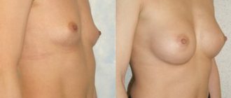
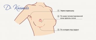
How is the operation performed?
- After introducing the patient into a state of general anesthesia, the surgeon makes an incision in the skin surrounding the nipple.
- After this, the skin is separated from the breast tissue, and the mammary gland is separated from the underlying muscles.
- Then comes the turn of lymphadenectomy, when the surgeon, using a scalpel or scissors, removes the tissue that contains the regional lymph nodes of the mammary gland. This is the most delicate moment of the operation, since the tissue also contains main veins and arteries. It is necessary to free them from fiber without damaging them. Also, during the removal of tissue, surgeons try to preserve the intercostal-brachial nerve (n. intercostobrachialis), which helps preserve the sensitivity of the posterior surface of the shoulder after surgery, as well as reduce the likelihood of developing late postmastectomy lymphostasis of the arm.
- After complete removal of the mammary gland in a block with tissue, the wound is inspected for bleeding, thorough hemostasis of the wound is carried out, and drains (special tubes) are installed, which serve to remove fluid accumulating from the wound.
- After the drains are installed, the wound is sutured, and the outer parts of the drains are connected to a vacuum tank, popularly nicknamed the “accordion” for its characteristic appearance.
As a result of the operation, a horizontal scar remains at the site of the mammary gland and nipple.
Symptoms of serous mastitis
The symptoms of the disease are quite common at first, and resemble a common cold, but after 2-3 days, mastitis becomes acute and its symptoms become quite specific.
- Increase in body temperature, sudden jumps to 39-40 degrees, followed by a decrease to normal temperature.
- Chills, fever and other common symptoms of inflammation.
- Redness of the skin at the site of inflammation.
- Poor milk flow, sometimes inability to continue feeding.
- Bursting pain in the mammary gland.
- Swelling of the gland, redness of the skin at the site of inflammation.
Serous mastitis begins like a regular flu; most women, experiencing malaise, weakness, and drowsiness, attribute the symptoms to a cold. A little later, specific symptoms are added to the familiar ones: chest pain, redness of the skin at the site of inflammation. Body temperature rises, breasts swell and feeding becomes difficult
It is important to recognize mastitis immediately and consult a doctor immediately. Signs of acute mastitis require immediate treatment
Redness of the skin is an external sign of an inflammatory process in the chest
Treatment of neoplasm
Inflammatory processes in the mammary glands must be eliminated using the following methods of influence:
- antibacterial drugs for oral administration;
- intravenous administration of antibiotics;
- use of complex vitamins;
- the use of medications aimed at strengthening the immune system.
In order to avoid inflammatory processes in the cyst, complex therapy is required. It includes the use of the following medications:
- painkillers. They are aimed at eliminating the unpleasant symptoms that accompany a breast cyst. The most effective means of this group are:
- Nimesil;
- Nurofen;
- Diclofenac.
- Antibacterial drugs. They help relieve inflammatory processes. Their use is especially useful during the rehabilitation period after surgical removal of a tumor. Examples of antibiotics:
- Cefepime;
- Ceftriaxone;
- And so on.
- Immunomodulating drugs. The vitamin complex is required to maintain the body in normal condition, as well as to effectively fight the cyst. To do this, you can use the following tools:
- Echinacea;
- Vitrum;
- Immunal.
Also, during the treatment period, sedative medications will be useful. Since the state of stress and emotional overstrain have an extremely negative impact on the course of this disease.
If the inflammatory process develops rapidly, the attending physician prescribes surgery. Surgery is required to remove the pathological tumor. The duration of the rehabilitation period is about 10 days.
This is how long it takes to remove the stitches. The prognosis for such an operation is usually favorable. After just 2 weeks, a woman can return to her normal, habitual lifestyle.
To prevent the recurrence of a cyst in the breast, a woman should follow a few simple rules:
- regularly visit a mammologist for preventive examinations;
- avoid injury to the mammary glands;
- during breastfeeding, avoid stagnation of milk;
- wear comfortable underwear made from natural fabrics;
- spend a minimum amount of time in direct sunlight.
You should also not forget about maintaining immunity.
It will be useful to familiarize yourself with the following materials:
- Why cysts form in the mammary glands: causes and diagnosis of the disease.
- Reasons for the appearance of a cyst in a woman over 40: what to do for preventive purposes?
- What causes cysts to appear in teenage girls and other children?
- What are the signs and symptoms of breast cysts in women?
- What is a ductal cyst of the mammary glands?
- Why do cysts in the mammary glands hurt and when should you see a doctor?
- A proper diet for breast cysts is the key to a speedy recovery!
- Neoplasms in the mammary glands: how to distinguish a cyst from breast cancer?
- Is a breast cyst dangerous?
Symptoms
Serous mastitis is an unpleasant and dangerous disease for women. If it is diagnosed quickly, treatment will be quick. But if the disease is neglected, complications may develop. The main symptoms of serous mastitis in women:
- temperature increase;
- the mammary glands become hot to the touch;
- swelling of the breast, a feeling of weight;
- local redness of the skin;
- bad feeling.
Some inexperienced mothers may confuse this serious illness with ordinary lactostasis. But unlike it, with serous mastitis, after emptying the breasts, a woman does not experience a feeling of relief. In some cases, a new mother may have a problem with her milk flow, which can make breastfeeding difficult. Without treatment for serous mastitis, a woman experiences nausea, vomiting, and dizziness.
A young mother should pay increased attention to the condition of her nipples. If a woman notices that they are cracked, then she needs to make an appointment with a doctor
Upon examination, the doctor may notice enlarged lymph nodes in a sick young mother. The woman has chills and is weakened. In advanced cases, there is an abscess at the center of the inflammatory process on the mammary gland. In some cases, such formation may not be isolated.
Treatment of serous mastitis
To normalize blood circulation and improve lymph circulation, it is important to restore normal milk flow. After the swelling subsides, the serous fluid will be absorbed spontaneously, and the disease will be cured. To improve your milk flow, it is important to continue breastfeeding or learn proper pumping techniques. To facilitate this process, perform a light and gentle massage from the top of the breast to the nipple. The next massage movement is carried out from the nipple to the armpits along the flow of lymph.
To improve blood circulation and relax the chest muscles, you can place a warm heating pad on the mammary gland.
It is not necessary to use medications to treat serous mastitis.
Medicines for symptomatic treatment:
- No-spa – relieves pain, improves milk flow;
- Malavit is a medicine created on a natural basis, used for compresses that relieve pain and itching, swelling and inflammation;
- Progestogel is a gel with progesterone in its composition, used to relieve swelling and reduce vascular permeability;
- Menovazin is an ointment for topical use, used to restore blood circulation and relieve swelling.
Vitamin complexes are used to restore immunity.
Physiotherapeutic methods are used in the treatment of serous mastitis:
- Electrophoresis with magnesia, Dimexide, Malavit;
- Infrasound;
- Magnetotherapy.
Proven folk methods are used as additional means. Among them are compresses made from honey cakes and cabbage leaves, alcohol compresses with aloe juice. It is important to remember about the dangers of alcohol for an infant and to wash off traces of such a compress from the breast.
Medicinal herbs are used as compresses, ingredients of ointments and healing infusions:
- Arnica – infusion relieves swelling, helps resolve infiltrate, improves blood circulation.
- Yarrow - compresses from brewed herbs or using an infusion internally helps relieve spasm of the milk ducts and normalize lactation.
- St. John's wort – compresses have an antispasmodic effect and improve blood flow from the source of inflammation.
Causes
Awareness of the causes that provoke the development of serous mastitis will allow you to avoid it through prevention. This is an inflammation of the mammary glands, in which the process of suppuration has not yet begun. If the disease appears, it is better to carry out therapy at the initial stage so that it does not turn into purulent mastitis.
Inappropriate breast care often provokes the development of serous mastitis. Maternity hospital specialists explain how to do this correctly, so all important points must be observed. There is no need to wash your breasts before and after each breastfeeding. Rubbing your nipples with a drop of milk will suffice. When carrying out such manipulations, there is no need to vigorously rub the nipple to prevent cracks from appearing. For hygiene purposes, it is enough to take a shower in the morning. The fewer interventions, the more successful breastfeeding will be.
During feeding, the baby should grasp the nipple correctly, not pull it and eat food calmly. In this way, it will be possible to avoid the appearance of cracks, which can provoke serous mastitis. The disease develops when the full outflow of milk is disrupted and stagnation begins. Breast milk creates a breeding ground for pathogenic microorganisms and bacteria. The outflow of intercellular fluid is also disrupted, as a result of which the tissues of the mammary glands with serous contents become inflamed. The penetration of bacteria through damaged skin into the serous infiltrate contributes to the development of inflammation and the appearance of purulent mastitis.
Serous mastitis can also occur in women who do not breastfeed. The most common cause of this phenomenon is surgical intervention on the mammary gland. If a woman has resorted to the services of a plastic surgeon, increasing the size of her breasts, the anatomical component of the mammary glands, which are compressed by the implant, may be disrupted. This picture provokes the onset of artificial lactostasis, leading to the appearance of mastitis.
Pathological causes of breast enlargement
Swelling, swelling and tenderness of the mammary glands can be a symptom of the following diseases:
- Lactostasis (blockage of the mammary gland duct with stagnation of milk). In the absence of therapy, it can serve as the beginning of an inflammatory process (mastitis). With proper feeding and expressing milk, it can go away on its own.
- Purulent mastitis. The inflammatory process in the mammary gland with the formation of foci with purulent contents is characterized by severe swelling and redness of the breast, the presence of purulent discharge from the nipples, high fever and signs of general intoxication of the body. In this case, surgical treatment to remove purulent contents and anti-inflammatory therapy are required.
- Benign neoplasms of the breast. Mastopathy is a pathological growth of gland tissue due to hormonal disorders.
- Fibroadenoma occurs against the background of fibrous mastopathy.
- A breast cyst is a collection of fluid that occurs when a breast duct is blocked. In the absence of inflammation it does not cause concern; in an inflamed state it can lead to swelling, redness and pain in the chest.
Symptoms of lactation mastitis
The first manifestation of lactostasis and lactation mastitis is pain (mastalgia) and/or the sudden appearance of focal lumps in the mammary gland during feeding.
The mechanism of occurrence of such painful sensations differs from other pains. This is due to the peculiarities of the sensitive innervation (connection with the central nervous system) of the mammary glands, namely the absence of pain receptors in them.
During breast surgery (those performed under local anesthesia), you do not even need to inject an anesthetic solution into the tissue itself. It is injected only into the skin and capsule of the gland, and the patient feels the gland itself weakly, even if she works with the parenchyma with a surgical instrument. Hence the conclusion that all pain in the chest is associated with an effect on the gland capsule: mechanical - during stretching, chemical - after the onset of inflammation. In other words, pain can only occur when lactostasis stretches the capsule. If this does not happen, then there may be no pain syndrome.
With mastitis, pain also appears due to stretching of the capsule or due to the impact of aggressive inflammatory mediators on it.
Soreness and thickening in the mammary gland can be accompanied by an increase in body temperature due to the action of bacterial breakdown products entering the bloodstream. This in itself is not a sign of mastitis. At this stage, you should stop feeding the child with the diseased gland and try to immediately carry out measures to express milk: by manual massage or using a special breast pump.
If intensive pumping does not bring relief within 2-3 days or severe pain syndrome no longer allows you to work with the gland, you should consult a doctor. The specialist’s task will first of all be to make the correct diagnosis: is it still lactostasis or is it already mastitis. Unresolved lactostasis most often develops into lactation mastitis 3-4 days after the onset of the disease. If the patient notices redness of the skin over the area of compaction and/or pain, then inflammation has begun. In this case, there is more clinical evidence for incipient mastitis.
How to avoid lymphostasis after mastectomy?
During the operation, in addition to glandular and muscle tissue, lymph nodes and vessels are removed, resulting in the penetration of soft tissue. This is why lymph flows for a long time after a mastectomy.
Stagnation of exudate can last for several weeks or several months (lymphedema).
Doctors recommend early prevention to avoid the unpleasant consequences of surgery.
Measures to prevent this disease include the following:
- Wearing a bandage on the chest and a compression sleeve on the operated side;
- Application of special lymphatic drainage and general tonic massage;
- Physical therapy classes and general physical activity;
- The use of diuretic and metabolic drugs to accelerate fluid outflow;
- Rubbing special creams and ointments into the skin to relieve swelling.
Thanks to simple actions, it is possible to avoid unpleasant consequences and improve the overall health of the patient.
Diagnostics
To confirm the diagnosis and exclude other diseases, it is not always enough to analyze external signs.
Additionally carried out:
- Urine and blood analysis.
- Measurement of general temperature.
- Measurement of pulse, breathing rate.
- Palpation, inspection, checking the udder. The temperature of the lobes is measured, their density, size, presence of wounds, abrasions are checked, and color is determined.
- Examination for the presence of inflammation in the lymph nodes.
- Feeling, examining the nipples along the entire length;
- Microflora research.
Milk testing
One of the key diagnostic methods is milk bioanalysis.
There are several ways:
- With Mastidine solution. Take 1 ml of milk and Mastidine (10%), stir with a wooden stick, leave for 15 minutes. If flakes appear in the mixture, it turns orange and takes on the appearance of jelly, the test is positive. Determines the level of acidity and the number of leukocytes.
- Using control plates with black and white indentations. Milk is dripped into the holes. In some cases, Mastidine (2%) is added. As an alternative, Dimastin (5%) is used. The flakes are clearly visible in the dark depressions; blood is visible in the white ones.
- Bromothymol test. Bromothymol solution (0.5%) is added to 1 ml of milk. The use of distilled water is allowed. If the mixture turns blue, yellow or bright green, it is a sign of inflammation.
- Measuring electrical conductivity using the Mastiton device. In a sick cow, the indicator is more than 600, in an animal at risk - from 450 to 600. The test is carried out many times. Milk for diagnostics is taken from all areas. There is a high probability of error; the results are double-checked with other tests.
- Advocacy. For this method, milk is taken from all parts, poured into a separate vessel, and placed in the refrigerator. Let it sit for 17 hours, then take it out, put it in the light, check the sediment, color change, and width of the cream. The test is positive if the milk is watery, the layer of cream is less than 5 mm. The method is considered unreliable.
Symptoms of non-lactation mastitis
The clinical picture of the disease is represented by local manifestations of the inflammatory reaction, pain and symptoms of general intoxication. The key feature of mastitis not associated with lactation is the less severe clinical manifestations. The disease, as a rule, debuts acutely with pain in the affected mammary gland, hyperthermia up to +37.5-38.0 ° C, and redness of the skin. The breasts look somewhat enlarged and swollen, and feel denser to the touch. In the presence of wound damage, pathological changes are more pronounced in the wound area; in other cases, they are usually widespread and diffuse. Patients often complain of weakness, chills, and headache.
After 1-3 days, the disease enters the infiltrative phase, as evidenced by the appearance of local compaction of breast tissue with clear boundaries, increased pain and hyperemia of the skin. The temperature continues to rise, reaching + 38.5° C or more. Intoxication also intensifies, the woman feels chills, fatigue, reports sleep disturbances, decreased or complete absence of appetite. Already at this stage, the axillary lymph nodes may enlarge. The duration of the period of infiltrative changes usually does not exceed 5-7 days, after which, in the absence of adequate treatment, tissue suppuration occurs.
At the purulent stage of the disease, one, several or many cavities filled with pus form in the damaged areas. The skin over the inflamed area appears bright red, and the pain becomes throbbing. As the temperature rises to + 39° C or higher, nausea and vomiting are possible. In some patients, even the purulent version of mastitis occurs mildly, with moderate pain and low-grade fever. In such cases, the inflammatory process is likely to become chronic with periodic exacerbations, deformation of the mammary gland, the formation of purulent fistulas and suppuration from the nipples during relapse.
Symptoms
Timely diagnosis and treatment of lymph accumulations in the breast tissue will protect against unpleasant consequences. It is for this reason that it is necessary to regularly consult with a specialist and pay attention to any discomfort in the chest area.
Symptoms of seroma after breast surgery:
- Redness - the color of the skin at the site of fluid concentration can range from yellow to scarlet due to the pressure exerted by the fluid on the vessels, which subsequently begin to burst.
- Deformation of the mammary gland - swelling may appear in the area of lymphoma accumulation, as well as enlargement of the gland, changes in its contours, and displacement of the nipple.
- The serous substance penetrates the wound - this complication occurs quite often in women with thin skin. A fistula may even form on the surface of the skin, through which the fluid will escape.
- Swelling of the breast tissue - fluid may well penetrate into the soft tissue of the breast, which is why in some places there is excessive elasticity of the skin.
- Painful sensations – pain with seroma is insignificant, but may increase with palpation, walking or physical activity.
If at least one of the above signs is noticed, you must immediately inform your doctor. Carrying out diagnostics and timely initiation of treatment allows you to eliminate the problem quickly enough.