Determining the age of skin scars
Prescription of scars (K.I. Khizhnyakova, 1945)[1]
| Duration of the scar | Capillaroscopic picture |
| up to 2 weeks | The background of the scar tissue is clear, pale pink. The vessels are in the development stage, extend from the edges of the scar, but do not reach the center and do not anastomose. The center of the scar is pale. There is no pigmentation of the scar or surrounding tissue. |
| 2 weeks–1 month | The background is pink. The pale central part of the scar narrows. The vessels at the edges come closer to each other. No pigmentation |
| 1-2 months | The background is uniformly reddish-bluish, cloudy, the vessels are dilated, go through the entire scar, anastomose, their number increases. |
| 2-6 months | The bluishness of the background gradually decreases, the background becomes clearer, acquires a pinkish color, and scar pigmentation appears. The number of vessels gradually decreases |
| 6-12 months | The background is pale pink, clear, later brown, sometimes heterogeneous. The number of vessels decreases, they become narrow. Small folds appear on the surface of the scar. Individual large vessels are visible. |
| more than 1 year | The background is grayish-white or pale brown, sometimes heterogeneous. There are few vessels, they are located in nests. The capillaries of the skin surrounding the scar are inclined towards the scar and are narrow. |
Duration of scars (I.M. Serebrennikov, 1962)[2]
| Duration of the scar | Properties of scar | ||
| color and shades | density | other signs | |
| up to 1 month | Pinkish, later reddish with a bluish tint | Soft | Flat, tender, crusty |
| 1-2 months | Reddish, with various shades of purple, usually dark purple | Dense | Convex, sedentary |
| 2-3 months | Reddish. The blueness gradually decreases. | Dense throughout | Convex, hypertrophic in nature |
| 3-6 months | The blueness disappears. Pink color begins to predominate | Gradually softens | Convex, sometimes retracted or level |
| 0.5-1.5 years | Pale pink. A brown color of various shades appears. Later whitish, with isolated areas of brown | Slightly dense or soft. Scar density varies | The surface is uneven or smooth, at or below the surrounding tissue |
| Over 1.5 years | More often whitish (white), less often – brown | Soft, dense cords or dense throughout | Thin, atrophic, shiny, sometimes convex |
“... The color and density of scar tissue have long been used as criteria for judging the time of occurrence of skin scars. Thus, E. Pelikan (1875) pointed out that for an approximate judgment about the time of castration, it is important to take into account the appearance, color and density of scars. Brief information on the issue of determining the prescription of the GGB is available from Kornfeld (1885), N.A. Obolonsko) (1894), Wieber (1908), Ottolenghi (1910), Smith ...., 1943), Kerr (Kegg, 1946), etc. These authors note that the age of scars can only be determined very tentatively, within a few months from the moment the scars appeared.
We find the most complete information on the issue of determining the age of scars in K. I. Khizhnyakova (1945) [1]. She suggests using a number of signs when solving this issue: color, density, the nature of the surface of the scars, their edges and the degree of mobility of the scars. When using the color of scars as a criterion for their age, it is necessary to take into account several conditions: the localization of scars, their massiveness, the possibility of accidental contamination, etc. Based on his own observations of scars, mainly of gunshot origin, K.I. Khizhnyakova provides indicative information for determining the age of scars by color.
As the author notes, these data cannot be fully applied to facial scars, since due to the abundant blood supply to this area, the red color of the scars lasts much longer. K.I. Khizhnyakova notes that the density of the scar mainly depends on its age, however, here too it is necessary to take into account the localization, extent, massiveness of the scars, the method and period of healing of the damage. The approximate timing of changes in scar density depending on their age is given. Over time, the scars (after 6-8 months) become soft and subsequently their consistency does not change. K.I. Khizhnyakova comes to the conclusion that the age of scars after gunshot injuries can be determined approximately within 1.5 years.
CM. Sidorov (1954)[3] examined 84 patients with gunshot wounds to resolve the issue of how long ago the wounds were. He believes that scars up to 2 months old are soft, from 2-3 months to 4-6 months they are slightly dense, and after 6-12 months they are dense. K.I. Khizhnyakova, who studied a significant number of scars after gunshot wounds (bullet and shrapnel), came to the conclusion that the greatest density of scars is observed up to 3 months, and then it gradually decreases (from 3 to 6 months) and after 6-8 months the scars become soft.
It should be noted that in small scars, the formation of which ends much sooner, the color change occurs faster than indicated by I.M. Serebrennikov. Scars that appear at the site of skin damage from the cutting of surgical sutures quickly become white and hardly noticeable, while in the main scar the process of its formation and corresponding color changes take place for a long time. The color change is due to the general formation of scar tissue. The faster the formation occurs, the faster the color changes. In very extensive scars, color changes may occur more slowly than indicated by I.M. Serebrennikov.
A known influence on the color of the scar is its localization. Scars located in the face and lower extremities, due to the peculiarities of blood circulation, retain a bluish tint for a very long time. Changes in scar density occur, judging by our data, as follows. Fresh tripe usually has a soft consistency. Around the end of the 1st month, the volume of scar tissue increases, the scar begins to protrude above the skin level and evenly thickens, becoming dense. Scars can remain in this state for various periods of time - until the end of the 2nd or even 3rd month. Older scars gradually soften and flatten. In such scars, all these cyclic changes that are normal for a scar are poorly expressed and it is often not possible to notice changes in density by touch.
It should be emphasized that the density and nature of scars depend not only on their age, but also on the amount of fibrous tissue formed during the formation of the scar, the depth of the former damage and localization. In massive scars of considerable length, the density of scar tissue throughout the scar is often heterogeneous. Often scars, having become dense, remain in this state for many years. In some cases, this is due to hyalinosis of scar tissue. The conclusion about the age of scars is based on such objective data as the color, density and nature of the scar tissue. Capillaroscopic examination of them can also help determine the age of the scar. In addition, you need to take into account various medical and investigative documents, which often indicate the time of receipt of certain injuries. The expert must also critically take into account such subjective data as the testimony of the examinees about the time of scar formation. Often he has the opportunity, without establishing the exact age of the scars, to indicate in the conclusion that they appeared at different times. And such a conclusion can be very useful for investigative authorities. It is impossible to recognize as scientifically substantiated the still encountered conclusions of forensic experts, which accurately indicate the age of scars after the end of their formation (for example, “the age of the scar is 5 years, 10 years,” etc.). Based on the study of only the external properties of the scar, such conclusions should be considered erroneous and scientifically unfounded.” [2]
Hypertrophic and keloid scars
Scars are the result of replacement of damaged own tissues with rough connective tissue as a result of surgical interventions and various traumatic factors (mechanical, temperature, chemical, ionizing radiation, deep destructive inflammation, etc.) [1, 2, 3]. Scars, being a pronounced cosmetic defect, often lead to psycho-emotional discomfort, as well as the development of psychosocial maladjustment and a decrease in quality of life.
Scar formation goes through several stages:
- Stage 1 - inflammation and epithelization on days 7–10 after injury. The edges of the wound are connected by fragile granulation tissue; there is no scar as such yet. This period is very important for the formation of a thin and elastic scar - it is necessary to prevent suppuration and divergence of the edges of the wound.
- Stage 2 - formation of a young scar. This is 10–30 days after the injury. Collagen and elastin fibers begin to form in the granulation tissue. Increased blood supply to the injury remains - the scar is deep pink in color.
- Stage 3 - formation of a “mature” scar lasting from 1 to 3 months after injury, blood vessels completely disappear, collagen fibers line up along the lines of greatest tension. The scar becomes light and dense.
- Stage 4 - final transformation of the scar. Duration 4–12 months after injury.
Scar formation occurs mainly due to the extracellular matrix, in particular with the help of collagen. The extracellular matrix is a supromolecular complex that includes chemical compounds of various types (proteins, polysaccharides, proteoglycans, and others). It can be compared to a gel in which fibrous proteins (collagens and elastin) and viscous proteins (fibronectin, laminin) “float”, ensuring the interaction of molecules with each other and with cell surfaces [2, 9, 14, 17]. Of all proteins, collagens constitute the main component of the extracellular matrix and are the most abundant proteins in the body, accounting for about 1/3 of all its proteins. These are water-insoluble extracellular glycoproteins synthesized by fibroblasts, chondroblasts and osteoblasts. Due to the shape of the protein molecule, collagens are classified as fibrillar proteins [16]. The growth of excess extracellular matrix in the scar occurs as a result of the activity of “wound” fibroblasts. In intact (healthy) skin, fibroblasts are responsible for remodeling the components of the dermis; they destroy old collagen and lay down new one. During wounds, injuries, burns and surgical interventions, myofibroblasts appear in the wounds, which strive to “close the gap” in the tissues, intensively depositing components of the extracellular matrix: collagen, glycosaminoglycans, elastin and other proteins. It is due to the proliferation of fibroblasts and their production of excess extracellular matrix that scar growth occurs [2,19].
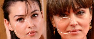
The following types of scars can be distinguished: normotrophic, hypertrophic and atrophic. During a normal physiological process, a normotrophic scar is formed. These are flat, light-colored scars with normal or reduced sensitivity and elasticity close to normal tissue. Such scars are optimal. In a typical wound, 6–8 weeks after injury, a balance between anabolic and catabolic processes is established. At this stage, the strength of the wound is approximately 30–40% of the strength of healthy skin. As the scar develops, its tensile strength increases as a result of the progressive bonds of collagen fibers.
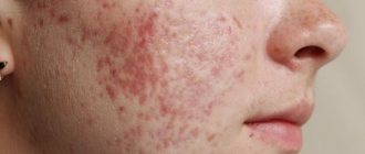
When the connective tissue reacts excessively to injury against the background of unfavorable healing conditions (inflammation, scar stretching, etc.), hypertrophic scars are formed. Under conditions of hypoxia and inflammation, fibroblasts are activated by biologically active substances. Undifferentiated, pathological, functionally active cells with a high level of collagen synthesis appear. The formation of collagen prevails over its breakdown due to a decrease in the production of collagenase, a specific enzyme that destroys collagen, as a result of which powerful tissue fibrosis develops in the form of hypertrophic or keloid scars. Hypertrophic scars are often combined into a group with keloid scars due to the fact that both types are characterized by excessive formation of fibrous tissue and arise as a result of inadequate inflammation, the addition of a secondary infection, a decrease in local immunological reactions, etc. However, there are also significant differences, such as: on the basis of which differential diagnosis is carried out between these two types of scars, which will be mentioned below [3].
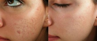
Atrophic scars result from a decreased response of connective tissue to injury. Not enough collagen is formed. Atrophic scars are located below the level of the surrounding skin (sink). With a small width, they practically do not differ from normotrophic ones. Often, atrophic scars appear in places of former exfoliation (acne) - the so-called “post-acne”.
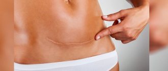
Keloid (keloidum: Greek kele bulging, swelling + eidos appearance; synonym: Alibera keloid) is a dense growth of connective tissue of the skin, reminiscent of a tumor [2]. Keloid scars develop as a result of a perverted tissue reaction to injury; this is a special, most severe group of scars that differ from others in appearance and pathogenesis. As a rule, keloids form against the background of reduced levels of general and tissue immunity. When examining keloid tissue, extremely active fibroblasts are found, the degree of their activity is 4 times higher than that of cells during the normal healing process [9]. Collagen (mostly immature) is located in the form of wide loose bundles and nodes, elastin is absent. A keloid scar has an elastic consistency, an uneven, slightly wrinkled surface. Keloid is usually classified into true and false. A true (spontaneous) keloid scar occurs spontaneously without any prerequisites, most often on the skin of the chest, along the outer surface of the upper third of the shoulder, and in rare cases throughout the body. A false keloid can develop in any place where there has been previous trauma to the skin (mechanical, burn, etc.). For example, a casuistic case was observed by Chawla B., Agarwal A., Kashyap S. (2007), who diagnosed a keloid formation on the cornea of the eye after surgery. Currently, this classification is relative. Most researchers believe that spontaneous keloid is preceded by microtrauma to the skin and etiologically it is no different from a false one (in pathogenesis and morphology, these scars are absolutely identical) [2]. A keloid at the site of abrasions or scratches is a raised area. The color and intensity of the color depends on the degree of vascularization (bright pink, pale, cyanotic). A keloid scar is characterized by pulsating growth, like the ebb and flow of the tide. Scar tissue in keloids extends beyond the original wound, usually does not regress spontaneously, and tends to recur after excision.
Theoretically, keloid can appear at any age, but most often develops during and after puberty. Young individuals are more likely to be injured, and young skin creates more tension, while older skin is less elastic and more rigid. Collagen synthesis levels are also higher at younger ages. Currently, the average age of patients is 25.8 (22.3 in women and 22.6 in men) [5].
Typically, diagnosing a keloid is not difficult. Gulamhuseinwala N., Mackey S., Meagher P. (2008), who conducted a retrospective study of 568 patients with keloids, found that the clinical diagnosis coincided with the histological diagnosis in 94% of cases. However, when scars begin to appear, the question of differential diagnosis between hypertrophic scar and keloid arises. As mentioned above, there are significant differences between these types of scars. The growth of a hypertrophic scar begins immediately after healing and is characterized by the formation of “plus tissue”, an area equal to the wound surface, while the boundaries of the keloid always extend beyond the damaged area. With a hypertrophic scar, subjective sensations are absent or insignificant. Keloid formations cause various subjective sensations (itching, pain, feeling of skin tightness, parasthesia, etc.). The change in color of a hypertrophic scar from pink to whitish occurs at the same time as in normotrophic scars, and over time they noticeably flatten. The keloid remains deep in color and does not regress spontaneously. Typically, one patient has several keloid formations. Keloid scars are most common in people aged 10–30 years, while hypertrophic scars can occur at any age. The morphological picture also has significant differences. It is known that collagen synthesis in keloids is approximately 8 times higher than in hypertrophic scars, which explains the lower quantitative content of collagen fibers in hypertrophic scars, and therefore the mass of the scar. In hypertrophic scars there are fewer fibroplastic cells than in keloids, and there are no giant, immature forms of the “growth zone” [1, 7, 8, 9, 12, 21].
Researchers have noted an unequal frequency of localization of keloid formation in individuals of different races. For example, in white-skinned people, keloids often form on the face (cheek and ear), upper extremities and sternal area; and in Asians the sternal region is almost not affected; in blacks, along with the auricle and neck, the lower extremities are more often affected [5]. This suggested the leading role of heredity in keloid formation. However, to date, no specific gene or set of genes has been identified as being responsible for keloid development [5, 8]. In addition, no significant differences were found between keloid DNA fibroblasts and normal skin DNA fibroblasts. This suggests that what is important in the development of keloid formation is not the increase in the number of fibroblasts producing normal amounts of collagen, but rather that each fibroblast produces more collagen than is necessary for wound repair [13].
Treatment of keloid and hypertrophic scars
The first rule in the treatment of keloids and hypertrophic scars is their prevention. First, unnecessary cosmetic surgery should be avoided in patients prone to keloid formation and severe scarring. A possible exception would be a disfiguring keloid on the pinna. If surgery cannot be avoided, closure of all surgical wounds should occur with minimal tension along the skin folds whenever possible. It is advisable that the incisions do not pass through the surface of the joints and in the middle part of the chest.
Treatment methods for keloid and hypertrophic scars can be divided into several groups:
- medications (corticosteroids, immunomodulators, drugs affecting collagen formation);
- physical and physiotherapeutic (use of occlusive dressings and compression therapy, excision, cryosurgery, laser therapy, electrophoresis, etc.);
- radiation therapy;
- cosmetic procedures aimed at external correction of the defect.
Medicines
Corticosteroids. Intraruminal steroids remain the mainstay of treatment. Corticosteroids reduce scar formation by reducing the synthesis of collagen, glycosaminoglycans, inflammatory mediators, and fibroblast proliferation during wound healing. The most commonly used corticosteroid is triamcinolone acetate at a concentration of 10–40 mg/mL, administered into the injured area by needle injection at 4–6 week intervals. The effectiveness of such an introduction as a monomodel and as an addition to the scar excision procedure is very high. Topical corticosteroids are also widely used, which are applied daily directly to the formation. Complications of corticosteroid treatment include atrophy, telangiectasias, and pigmentation disorders.
Immunomodulators. A new method in the treatment of keloid and hypertrophic scars is interferon therapy. Interferon injected into the suture line after excision of a keloid scar can prophylactically prevent relapses. It is recommended to administer 0.5–1.0 million IU every other day for 2–3 weeks, then 0.1–0.5 million IU 1–2 times a week for three months.
Drugs that reduce hyperproliferation of connective tissue cells. The classic treatment for scars is hyaluronidase. Hyaluronidase breaks down the main component of the interstitial substance of connective tissue - hyaluronic acid, which is a cementing substance of connective tissue, and thus increases tissue and vascular permeability, facilitates the movement of fluids in the interstitial spaces; reduces tissue swelling, softens and flattens scars, and prevents their formation. Preparations containing hyaluronidase (Lidase and Ronidase) are obtained from the testes of cattle. Lidase solution (1 ml) is administered in these cases near the site of the lesion under the skin or under scar tissue. Injections are made daily or every other day; the course of treatment consists of 6–10–15 or more injections. If necessary, repeat courses are carried out at intervals of 1.5–2 months.
A new enzyme preparation for the treatment of diseases accompanied by the growth of connective tissue is Longidaza. "Longidase" is a chemical compound of hyoluronidase with polyoxidonium. The combination of the enzymatic activity of hyaluronidase with the immunomodulatory, antioxidant and moderate anti-inflammatory properties of polyoxidonium provides a wide range of pharmacological properties. It is most effective to use the drug "Longidaza" by ultraphonophoresis or phonophoresis. For ultraphonophoresis, Longidase 3000 IU is diluted in 2–5 ml of gel for ultrasound therapy. The impact is carried out with a small ultrasonic emitter (1 cm2), with an ultrasound frequency of 1 MHz, intensity 0.2–0.4 W/cm2, in continuous mode, exposure time 5–7 minutes, course of 10–12 procedures daily or every other day . Using the phonophoresis method (1500 Hz), 3000 IU of Longidase is administered daily (total exposure time 5 minutes, course - 10 procedures). It is also possible to administer the drug inside the scar:
- small keloid and hypertrophic scars: Longidaza 3000 IU once every 7 days for a total course of 10 injections into the scar;
- keloid and hypertrophic large area of the lesion: Longidase 3000 IU 1 time in 7 days inside the scar in a course of 8-10 injections, at the same time intramuscular injection of Longidase 3000 IU No. 10.
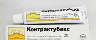
A well-known drug that inhibits the proliferation of connective tissue cells and at the same time has an anti-inflammatory effect is Contractubex. For many years, Contractubex has been used in surgery and cosmetology in the treatment of postoperative and post-burn scars, including rough scars that impede movement and keloids, as well as stretch marks (striae) after childbirth or after sudden weight loss.
The enzyme preparation of 9 collagenolytic proteases “Fermenkol” is a fundamentally new proteolytic drug. The anti-scar effect of Fermenkol is based on the reduction of excess extracellular matrix in scar tissue. The effect when using anti-scarring agents is observed approximately 3 weeks after the start of use of the product and the optimal result is usually achieved after 2-3 courses of electrophoresis or phonophoresis, 10-15 sessions or applications for 30-60 days.
Physical and physiotherapeutic methods
Silicone sheets and silicone occlusive dressings have been used with varying degrees of success in the treatment of hypertrophic scars and keloids. When using silicone dressings, the effect is more a result of a combination of pressure and surface hydration than the effect of the silicone itself. Studies show that the effectiveness of such dressings when used 24 hours a day for 12 months was observed in only ≈1/3 of patients [20]. The compression/pressure method achieved with silicone sheets causes thinning of the skin. In one study, applying two plates to the earlobes after keloid removal prevented its recurrence for 8 months to 4 years. A significant discomfort of the method is the need to wear bandages/plates throughout the day, as well as the impossibility of attaching them in some anatomical areas (folds, neck, etc.). Other devices (special compression garments and zinc oxide patches) are also used 12 to 24 hours a day for 8 to 12 months, which is a clear disadvantage of the method. According to various authors, their effectiveness does not exceed 40% [4, 5].
Cryosurgery. Cryosurgical agents, such as liquid nitrogen, attack the microvasculature and cause cell death through the formation of intracellular crystals. Typically, 1–3 freeze-thaw cycles of 10–30 seconds are sufficient to achieve the desired effect. Particular attention should be paid to the short duration of liquid nitrogen applications to avoid the appearance of reversible hypopigmentation. Please note that cryotherapy can be painful and cause depigmentation. A more promising method is the combined effect of cryotherapy and corticosteroids [15]. A simple and effective method of facilitating intraruminal steroid injection is to apply very light cryotherapy prior to injection, a step that will cause tissue swelling and subsequent cellular and collagen destruction. Liquid nitrogen is applied for 5-15 seconds, without allowing the skin to freeze. This technique allows for better distribution of the drug in the keloid tissue and minimizes its penetration into surrounding tissues.
Laser therapy. Argon laser (488 nm) is similar to carbon dioxide laser and can promote collagen contraction through local heating. A pulsed dye laser (585 nm) causes a process of photothermolysis, leading to microvascular thrombosis. In the early 1980s, authors noted that with the use of pulsed dye laser, scars became less erythematous, softer, and less hypertrophic. Laser treatment of keloids and hypertrophic scars using a carbon dioxide laser (10,600 nm) can ablate (can cut and cauterize) the lesion, creating a dry surgical environment with minimal tissue trauma. When carbon dioxide laser is used as a single model, the recurrence rate of keloid is quite high, so it is recommended to combine laser treatment with postoperative steroid administration, which significantly reduces the recurrence rate.
Radiation therapy. The use of radiation therapy to treat keloids remains controversial. One retrospective study found that the use of superficial radiation therapy after scar resection caused recurrence in 53% of patients [18]. Preoperative treatment of scars with a solution of hyaluronidase, followed by surgical removal and then external irradiation, is considered more promising to reduce the likelihood of relapse [6].
X-ray therapy (Bucchi rays) is based on the action of ionizing radiation on connective tissue, causing swelling and destruction of collagen fibers and fibroblasts. X-ray therapy is prescribed up to 6 irradiation sessions with an interval of 6–8 weeks at a single dose of up to 15,000 R. Only the superficial layers of the skin (in particular, the scar) are exposed to ionizing radiation, and the X-ray load on the underlying tissue is insignificant. Contraindications to Bucca therapy are kidney disease, circulatory decompensation, the presence of dermatitis and residual wounds. The idea of using x-ray therapy is very rational, because in the case of destruction of a certain number of fibroblasts, a balance is achieved between the synthesis and degradation of complex collagen, up to a change in its very structure. This idea, in particular, has been implemented using modern laser technology. The total radiation dose to prevent recurrence of a keloid is 15 to 20 Gy. They also recommend one-time irradiation of the wound on the day of suture removal in the same doses - 15–20 Gy. Sometimes this procedure is repeated up to 6 times at intervals of 1.5–2 months.
Excision. It is advisable to avoid surgery, especially if a keloid is present. Excision relapses in 45–100% of cases and should only rarely be used as a standalone model of therapy. After excision, long-term anti-relapse therapy is necessary. Whenever possible, wound pressure bandages and linens are used in the early postoperative period. Reducing relapses is achieved by combining excision with other methods, such as radiation or radiotherapy, interferon or corticosteroids [10].
After surgical interventions or skin injuries, it is necessary to take preventive measures to reduce the risk of keloid formation or hypertrophic sutures. The period of active therapy should begin at the 2nd stage of scar formation, i.e. 10–30 days after injury. According to the authors, there is a proportional dependence on the effectiveness of therapeutic treatment on the age of the scars: scars older than 6 months are less responsive to therapy than younger ones. Most likely, the decrease in the effectiveness of therapeutic measures is associated with a decrease in the number of vessels and compaction of scar tissue, which worsens its trophism [3]. Preventive methods may include: occlusive dressings, compression therapy, external applications or intradermal administration of glucocorticosteroids, Contractubex, Longidase, immunotherapy.
With a formed scar with a duration of up to 12 months, it is possible to carry out therapy with all methods, and with a long-term formation (more than 12 months), only aggressive methods are effective: injection of corticosteroids into the affected area, excision, radiation therapy, Bucca therapy, laser therapy.
Cosmetic procedures aimed at external correction of the defect. The most popular cosmetic procedures (peelings, mesotherapy, dermabrasion) do not serve any therapeutic purpose when affecting scars. The use of these methods contributes to the aesthetic correction of small scars. It is important to know that hypertrophic scars can be resurfaced only after they have reached a calm state. To obtain satisfactory results in the treatment of scars with a duration of more than six months, cosmetic procedures must be combined with 1–2 injections of corticosteroids (Diprospan) [2]. Sandblasting dermabrasion (10–15 sessions with an interval of 10–14 days) with a standard course of treatment provides satisfactory results, but when conducting two courses of 10–15 procedures, the effectiveness of treatment increases [2]. Surgical dermabrasion of hypertrophic scars requires re-operation after six months. Complete epithelization of hypertrophic scars after deep surgical dermabrasion occurs much later than epithelization of normotrophic and hypotrophic scars and lasts for 3–4 weeks, even when using wound healing ointments [2]. O. S. Ozerskaya (2002) performed a transplantation of autologous keratinocytes to prevent recurrence of scars after dermabrasion. Unhindered removal of the bandages is possible on days 7–8; complete epithalization of the scars occurred within up to 14 days. Thus, a significant acceleration of epithalization during dermabrasion of scars with keratinocyte transplantation is obvious. To avoid even greater hypertrophy and hyper- or depigmentation, hypertrophic scars cannot be corrected using aggressive methods, and medium peels can be done only after a course of superficial peels. As for keloid scars, it is better not to expose them to irritating methods, since there is a high probability of provoking their further growth.
Peeling with fruit acids. During peeling, dead skin cells are exfoliated and the regeneration processes of its deep layer are stimulated. Fruit acids gently penetrate the skin without damaging it and stimulate the production of collagen and elastin. Thanks to this, the elasticity of the skin increases, the relief is evened out and hyperpigmentation is reduced, which externally corrects the cosmetic defect of small scars.
Chemical peels. For small, mildly expressed hypertrophic scars, chemical peels are best performed in two stages. At the same time, in daily home care on the 21st day, in order to smooth out scar hypertrophy, a silicone dermatological cream is included, which creates an occlusive film, protecting the skin from moisture loss, relieves inflammation and erythema, and stimulates repair. In parallel with peelings, you can conduct mesotherapy sessions with regenerants and local microcirculation enhancers. Treatment of mildly expressed hypertrophied scars after a course of chemical peels can be continued with topical medications. For severe hypertrophic scars, peelings are carried out six to eight months after excision of the pathological scar or a course of physiotherapy (galvano-, phonophoresis) with hyaluronidase and heparin. The course of chemical peelings is also carried out in two stages: first, several sessions of multi-fruit peels (glycolic, salicylic, lactic, citric acids) once a week, then a complex retinol yellow peeling. Peeling is performed according to an individual scheme in accordance with the parameters of the scar and skin reaction.
In conclusion, it should be noted that the therapy of keloid and hypertrophic scars, with all the variety of treatment methods, requires a strictly individual approach, taking into account the main parameters of the scar: the size and duration of its existence.
For questions regarding literature, please contact the editor.
Yu. A. Gallyamova*, Doctor of Medical Sciences Z. Z. Kardashova** * RMAPO, ** Russian State Scientific Center, Moscow
Key words: hypertrophic scars, keloid scars, keloid disease, keloid, keloid formation, keloid treatment, scar treatment, scar prevention.
Buy an issue with this article in pdf