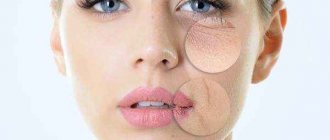
The appearance of a nasolacrimal groove is not necessarily due to aging, however, age-related changes in the architecture of the surrounding soft tissues entail an increase in its depth and severity. Dark circles and hollows under the eyes, a tired appearance are typical complaints that accompany the appearance of a nasolacrimal trough. Previously, chemical peels and surgical procedures were most often used to correct this cosmetic defect.
They have been replaced by less invasive techniques for replenishing volume with fillers, most often based on hyaluronic acid. Complications of contouring in the periorbital zone occur more often than in other areas of the face.
Therefore, knowledge of the anatomy of soft tissues and blood vessels , which will be discussed in the article estet-portal.com , is the basis for safe filling of nasolabial folds .
- Nasolacrimal, palpebromalar and midbuccal grooves
- What factors influence the formation and severity of the nasolacrimal groove?
- Anatomy of the nasolacrimal trough: muscles and ligaments
- Anatomy of the nasolacrimal trough: fat compartments
- Anatomy of the nasolacrimal trough: blood vessels
- Anatomy of the nasolacrimal trough: lymphatic vessels
- Recommendations for filling the nasolacrimal trough with fillers
Nasolacrimal, palpebromalar and midbuccal grooves
The nasolacrimal groove is the medial third of the depression that runs from the inner corner of the eye to the midline of the pupil in the infraorbital region. This structure consists of thin and low-elastic skin of the eyelid, as well as denser skin of the cheek. Along the nasolacrimal groove, the fascia is attached to the underlying periosteum.
The palpebromalar groove is located laterally. In patients with deep nasolacrimal and palpebromalar grooves, an almost continuous depression is formed, which is visualized several millimeters below the edge of the orbit.
The midbuccal groove is located below and medial to the nasolacrimal groove.
How to remove nasolacrimal trenches at home
Folk cosmetology products and grandmother’s proven methods will not remove nasolacrimal grooves forever; the effect will be short-lived and not always pronounced. Massages and gymnastics at home can be more effective and have a longer lasting effect. However, exercises need to be done every day, and massage in courses or also daily.
Let's consider all possible techniques and recommendations!
Folk remedies
Compresses
Compresses made from strong black or green tea will relieve morning puffiness around the eyes and the tired appearance of the eyes after a working day. Also, a compress made from chamomile decoction, which also relieves inflammation and soothes the skin, copes well with the same tasks.
A compress of cucumber juice with the addition of aloe juice (or without it) effectively refreshes the skin around the eyes and the area where the nasolacrimal groove forms.
Birch leaf compress. To do this, birch leaves are collected, filled with mineral water and infused for 12 hours.
Masks
- Potato mask. Boil the potatoes and mash them into a puree. Place it in gauze and apply it to the nasolacrimal grooves for 20 minutes. This mask has a pronounced rejuvenating effect.
- Cucumber mask. Grate the cucumber without the skin, add a little milk. Squeeze the resulting juice into a separate bowl, and put the thick mass into another container. First soak a cotton swab in the resulting juice. Take the thick mixture and spread it over the area around the eyes, covering with a moistened cotton swab. Keep this mask for 20 minutes. Be careful with your clothes because the juice will run down your face. It is advisable to be in a horizontal position at this time.
- Honey-yolk mask. Melt any honey and combine it with flour and egg yolk. Apply the mask to your face for 15 minutes.
Attention! If you want to say goodbye to nasolacrimal grooves at least temporarily with the help of masks, you need to do the masks every day until the effect appears.
Exercises for nasolacrimal trenches
- First, work the area above the wings of the nose. To do this, place your fingers under the eyes in the area of the upper cheeks. Make upward impulse movements with the lateral muscles of the wings of the nose.
- Contract the muscles of the inner corners of the eyes without wrinkling your nose. This exercise is aimed directly at pumping the nasolacrimal muscle. At the same time, while performing contractions, place your fingers in the area of the bridge of the nose and on the temples.
Nasolacrimal grooves, like other deep changes on the face, can be eliminated or their formation can be slowed down. Any of the methods described in this article requires both attention and capabilities. We advise you to first consult a cosmetologist who has sufficient reputation and experience. Then it will be possible to objectively assess the situation and build a competent plan of action aimed at getting rid of nasolacrimal grooves.
Tell us, what do you do to remove nasolacrimal grooves? Share your experience with other readers in the comments. Save the article in bookmarks so as not to lose it and come back to it again. Share it with your friends on social networks.
What factors influence the formation and severity of the nasolacrimal groove?
The formation of the nasolacrimal groove is influenced by:
- pigmentation;
- protrusion of fat packets;
- the appearance of wrinkles.
Possible causes of pronounced melanocytic pigmentation:
- excessive ultraviolet exposure;
- birthmarks;
- melanoma;
- dermal melanocytosis;
- diseases (systemic and skin);
- sleep disorders;
- lack of nutrients.
Dispigmentation does not affect the actual depth of the nasolacrimal groove, but increases it visually.
For hyperpigmentation in the infraorbital region, in addition to filling the nasolacrimal groove with fillers, additional correction methods may be needed.
The formation of a nasolacrimal trough is often associated with protrusion of orbital fat in the lower eyelid area. At the same time, fatty hernias of the lower eyelid pose a separate problem.
Orbital fat prolapse can be identified by the characteristic shape of the orbital fat compartments, which are often visible through the skin, with a central fat compartment shaped like a cigar.
Follow us on Instagram !
In patients with severe fat pad prolapse, dermal filler monotherapy may be contraindicated. An alternative in such cases may be lower blepharoplasty.
The appearance of wrinkles on the skin of the lower eyelid can also increase the severity of the nasolacrimal groove. Characteristic features of this zone:
- minimal amount of subcutaneous fat;
- thin skin;
- dynamism due to the activity of the orbicularis oculi muscle.
Prolapse of fat bags in the area of the nasolacrimal groove
Loss of volume in the central third of the face also increases the severity of the nasolacrimal groove due to:
- loss of the anterior projection of the upper jaw;
- prolapse and atrophy of malar fat pads.
Correction methods
It is impossible to get rid of the nasolacrimal groove by using external cosmetics. They are more suitable for the prevention of aging, and only to a very small extent in the early stages of its formation. Currently, surgical and conservative correction methods have been developed.
Surgical correction
It consists of using one of the methods for getting rid of nasolacrimal grooves:
- Surgical lipofilling, which is applicable mainly for young and middle-aged patients who are psychologically inclined only to the isolated elimination of an aesthetic defect without radical changes in the contours of their face. The essence of the operation is to first remove adipose tissue from other parts of the body (abdomen, thigh) and fill the area of the nasolacrimal fold with it under local anesthesia. This procedure is performed when there is low skin tone in this area by making an incision in the conjunctiva (transconjunctival blepharoplasty) and lasts about 40 minutes. In the presence of very pronounced age-related changes, access is made through a skin incision.
- The meaning of the second method is to separate the adipose tissue of the lower eyelids and then lower it under the skin in the area of the nasolacrimal groove. This technique is used as an addition to lower eyelid blepharoplasty or midface lift if the patient has lower eyelid hernias, a pronounced nasozygomatic groove, and many wrinkles and folds in the said area.
Surgical correction of the upper and lower eyelids
Surgical methods for correcting nasolacrimal grooves are quite effective and long-term. At the same time, they carry the risk of uneven filling of the nasolacrimal fold with fatty lumps with their subsequent contouring, especially in people with thin skin. In addition, performing the operations themselves under anesthesia or even under local anesthesia has a number of limitations associated with concomitant diseases.
How to get rid of a nasolacrimal trough - non-surgical methods
These include:
- Injection lipofilling.
- Mesotherapy and contouring.
- Hardware methods.
Injection methods
Injection lipofilling to get rid of nasolacrimal grooves is carried out using the adipose tissue of the recipient himself. It is obtained from the thigh, abdomen or other areas, centrifuged, washed and, using a syringe and cannula, injected under the skin in the area of the nasolacrimal groove.
Positive results last for 3 years, but often subsequently the adipose tissue can be contoured on the surface of the skin, creating an uneven relief. In addition, some of the fat cells do not take root and die, which also reduces the effectiveness of the procedure.
Read more about this method in the article “Lipofilling”.
Mesotherapy and contouring of nasolacrimal grooves using hyaluronic acid. If mesotherapy is advisable only for preventive purposes and at the very initial stages of fold formation, then correction of the nasolacrimal groove with hyaluronic acid fillers through contouring is the most effective non-surgical method.
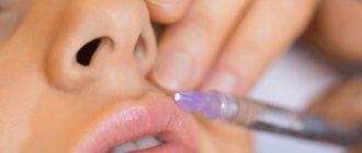
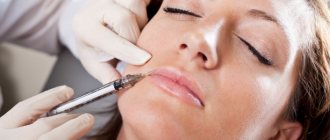
Application of filler
The method is relatively new and only in recent years has gained great popularity among both patients and doctors. This is explained by the almost absence of restrictions, the development of deep insertion techniques using a special cannula that prevents damage to blood vessels and nerves, as well as the emergence of the latest generation of fillers of not only high and medium, but also low density. In addition, the inclusion of lidocaine (a local anesthetic) makes the procedure painless and comfortable.
The different viscosities of the drugs allow them to be used selectively. For example, due to thin skin, only low-density hyaluronic acid fillers are suitable directly for the nasolacrimal groove, and for its branch, the nasozygomatic groove, medium or high-density fillers are needed. The only drawback of contour plastic surgery is the relatively short period of retention of the effect - on average 6-8 months, after which it is possible to repeat the procedure to remove the nasolacrimal grooves again.
The softlifting technique is effective for correcting this area.
Softlifting
Hardware methods
- exposure to high-frequency electromagnetic pulses (RF-lifting, Thermage), which stimulate an increase in the number of fibroblasts and the production of collagen and elastin;
- lifting through the deep impact of ultrasonic waves generated by the Uitera System apparatus on the muscular aponeurotic system (SMAS).
It is more advisable to use hardware methods of influencing the nasolacrimal trench not as independent methods of correcting the nasolacrimal trench, but in combination with mesotherapy or contouring using hyaluronic acid fillers.
Anatomy of the nasolacrimal trough: blood vessels
The ophthalmic artery (OA) is the main vessel providing blood supply to the orbit. This is the first branch of the internal carotid artery, which originates after exiting the cavernous sinus and enters the orbit through the optic canal. However, there are cases when the ophthalmic artery emerges from the middle meningeal branch of the external carotid artery and enters the orbit through the superior orbital fissure.
The vascular network of the periorbital zone is characterized by a high degree of individual variability.
Branches of GA can be divided into two groups:
- Orbital:
- lacrimal;
- supraorbital;
- anterior ethmoid;
- posterior ethmoid;
- internal artery of the eyelids;
- frontal (terminal branch);
- nasal (terminal branch).
- Ophthalmic:
- long eyelash;
- short eyelash;
- anterior ciliary;
- central retinal artery;
- muscular.
Only a small proportion of the blood supply to the orbit comes from the carotid artery (through its infraorbial branches) and the orbital branch of the middle meningeal artery.
Occlusion of the long and short anterior and posterior ciliary arteries can result in symptoms of anterior ischemic optic neuropathy, including vision loss .
The orbit is surrounded by an extensive network of vascular anastomoses. The literature describes in detail anastomoses between the branches of the internal carotid artery, as well as the temporal arteries and orbital arteries. Occlusion of any of them can lead to vision loss.
No mistakes
· Take good care of the skin of the periorbital area. If there is a tendency to fluid stagnation, drainage procedures are necessary. Good quality of the skin and its membranes, normal tone of the orbicularis oculi muscle is “insurance” against the deepening of the nasolacrimal groove.
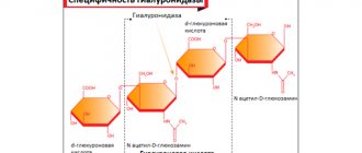
· A cosmetologist can lift and smooth out nasal tears strictly with filler based on hyaluronic acid! In case of introducing an excessive amount of the drug and overcorrection (the filler can be contoured in the form of a sausage), the doctor will use an antidote (hyaluronidase or longidase enzymes), which will break down the excess hyaluronic acid.
· If the filler is applied too superficially, the skin begins to glow blue. This complication is called the Tyndall effect. True, such a picture can be obtained with the correct injection technique - due to the individual characteristics of the tissue. Or because the correction was carried out earlier than three to six months after plastic surgery on the face, when complete recovery from the injury had not yet occurred.
· A filled nasolacrimal groove with an unrestored volume of tissue in the lateral orbit (the same palpebromalar groove) and the middle third of the face looks unaesthetic. Even if your focus is on a single flaw, complex aesthetic work is usually needed!
· It is preferable if the cosmetologist uses a cannula when correcting the nasolacrimal groove - this is the safest. When a doctor uses a needle, there is an increased risk of causing damage that can lead to embolism and blindness (fortunately, a rare complication). In this case, only super professionals can afford an igloo!
Anatomy of the nasolacrimal trough: lymphatic vessels
Lymphatic vessels of the periorbital zone are classified into pretarsal and posttarsal.
Lymphatic vessels of the periocular zone are divided into pre- and posttarsal. The outflow of lymph from the pretarsal part of the orbicularis oculi muscle and the skin of the eyelids is carried out through the pretarsal lymphatic vessels, and from the conjunctiva, lacrimal glands and tarsal plates - through the deep plexus of posttarsal vessels.
The outflow of lymph from the palpebral lymphatic vessels is carried out:
- laterally (into the deep and superficial parotid nodes) - from the lacrimal glands of the conjunctiva, lateral parts of the eyelids;
- medially (to the submandibular lymph nodes) - from the medial parts of the eyelids and lacrimal sacs.
True and false nasolacrimal trough
“With a true nasolacrimal groove, the tissue is tightly fused with the periosteum and there is practically no space for the introduction of filler,” notes our expert. “To smooth out such a groove, it is enough to strengthen the tissues of the periorbital zone with the help of mesotherapy and biorevitalization.”
Today there are drugs that give a good lifting effect, improve skin color, have powerful antioxidant properties, increase true collagen synthesis and even the production of elastin fibers!
A false nasolacrimal groove is formed due to ptosis—slipping of soft tissues, weakening of the ligament—and requires a different approach. Often this technique becomes an indirect correction - filling the missing volume of the cheekbone and mid-cheek area with fillers. “This is often enough to smooth out the nasolacrimal groove; the orbit begins to look very aesthetically pleasing,” says L. S. Babaeva. “If this effect is not enough, the filler is injected into the groove itself, so-called direct correction is carried out.”
Possible complications and methods of their treatment
The obtained result may differ significantly from the expected effect when correcting the nasolacrimal groove.
Here are some possible reasons for the deterioration:
- defective product;
- a cosmetologist who does not have the necessary knowledge and experience;
- violation of the conditions of the procedure;
- contraindications not detected during the examination.
All consequences are removed by performing certain manipulations when the client goes to a medical institution. In severe situations, surgical operations may be performed.
1. Swelling.
The appearance of swelling is a standard reaction of the body to various outside interventions. Normal swelling goes away on the second or third day. If one of the requirements is violated during correction, then the recovery process will change.
If fillers are injected too deeply or if the volume of the product is incorrectly selected, swelling may not subside within one to two weeks.
If, in addition to swelling, bruises begin to form on the skin around the eyes and not only, then an urgent consultation with a doctor is needed. Such a complication may be an indicator of damage to the circulatory system or signal the onset of inflammation under the skin.
Read material on the topic: Retinoic yellow peeling done by Hollywood stars
2. Pain.
Pain may persist for a long time when pressing on the area that has undergone correction. There are several explanations for this:
- nerve endings were affected;
- muscle tissue was shifted due to the administration of unequal doses of the drug;
- high sensitivity of the skin to the size of the needle used.
3. Inflammation.
Typically, inflammation is the body's reaction to the introduction of foreign substances. It usually goes away after two to three days. However, violation of sanitary and hygienic standards can lead to the development of infectious complications.
4. Necrosis.
Necrosis is the most dangerous and undesirable complication after correction of the nasolacrimal trough using fillers. Such changes develop if the administered drug gets between the vessels and disrupts the movement of blood in a certain area of the skin.
Swelling, inflammation, and bruising can cause necrosis. The gel, which has changed the movement of blood, itself begins to move and contributes to injury to the nerves. It is possible to eliminate the consequences and solve the problem of necrotic complications, but disturbances in the work of facial muscles are likely.
Nowadays, you no longer have to spend a lot of time performing complex and unpleasant procedures at home. It is much easier to seek help from real professionals - the Veronika Herba beauty and health center, equipped with effective and modern equipment.
Why clients choose Veronika Herba beauty and health center:
- This is a beauty center where you will have your nasolacrimal gland corrected at a reasonable cost, and your face will be treated not by an ordinary cosmetologist, but by one of the best specialists in Moscow. This is a completely different, higher level of service!
- You can receive qualified help at any time convenient for you. The beauty center is open from 9:00 to 21:00, seven days a week. The main thing is to agree with your doctor in advance on the date and time of your appointment.
Sign up for a consultation with a specialist by calling 8 (495) 995-15-13, and you will see for yourself!
Check-lifting ↑
The term Cheek-lift translates as “cheek lift”. The method tightens the middle third of the face and eliminates malar edema, pronounced nasolacrimal grooves, and eliminates bruises and bags under the eyes.
For plastic surgery, special endotin devices are used, which consist of polylactic and hyaluronic acids. They tighten and fix facial tissues in a new position, maintaining their elasticity.
Check-lifting
During the manipulation, one incision is made along the ciliary edge of the lower eyelid.
The plastic surgery takes from 1.5 to 2.5 hours.
Before and after rejuvenation with Cheek-lift
Filling the nasolacrimal trough: reviews, technique
At least 5-7 days before the procedure, it is recommended to discontinue the use of aspirin, NSAID drugs (Ibuprofen, Naproxen, etc.), vitamin E, and drugs with gingo biloba extract. This is necessary to reduce the risk of bleeding and bruising during or after the procedure. Some doctors, in the absence of contraindications, prescribe Dicynon tablets for the same purpose several days before the procedure. Great care should be taken in patients who have previously undergone lower eyelid blepharoplasty for tear trough correction.
It is safest to correct the nasolacrimal groove using a 25G cannula rather than a needle. Carrying out the procedure using a thin 30G needle is also possible, but is considered optimal only in patients after blepharoplasty, because in this case, there are many fibrous adhesions in the tissues, which will be difficult to penetrate with a cannula. It is optimal to use cannulas with a length of 38-40 mm, because the use of short cannulas is much more traumatic and more often leads to vascular complications.
The traditional insertion point for a 38 mm cannula is the intersection of the zygomatic and tragus lines. Anesthesia is required in the area of the injection point, and before anesthesia it is important to apply cold to the injection site, and as an anesthetic to use a drug containing a vasoconstrictor in a concentration of 1: 200,000 (a vasoconstrictor component such as epinephrine or others). Such rules for anesthesia will reduce the risk of bleeding and hematomas.
The classic correction technique is as follows: from the injection site, the cannula moves along the periosteum (i.e., under the orbicularis oculi muscle) to the inner corner of the eye, and during this process the tip of the cannula must be constantly palpated with a finger. The filler is released into the tissue only when the cannula moves backwards in a retrograde manner, and before starting removal it is extremely important to conduct an aspiration test. The injection volume on each side is from 0.2 to 0.4 ml, and will depend on the severity of the tear trough (24stoma.ru).
The upper border of the filler injection area is limited by the lower bony edge of the orbit (the place of attachment of the orbital septum) and, thus, in no case should filler be inserted above the lower bony edge of the orbit. The lower border of filler injection is the nasolacrimal ligament, which is located between the fibers of the orbicularis oculi muscle. Thus, the safe zone for filler injection will be only about 2-3 mm, and essentially a little higher or a little lower, or a little closer to the corner of the eye - literally means “shooting”. At the end of the procedure, it is important to gently massage the area with fingers.
Correction of the nasolacrimal trough with cannula: video
Advantages of using a cannula –
- minimum skin punctures.
- less painful procedure,
- less risk of hematomas and edema,
- there is less risk of embolism or occlusion of the angular artery, which can lead to necrosis of the skin of the infraorbital region and irreversible changes in vision.
The advantages of working with a needle are that correction, even with a very thin 30G needle, carries a significantly greater risk of vascular complications. However, this method also has its advantages, including...
- more accurate dosing of filler volume,
- more precise control of needle tip depth,
- optimal for patients after blepharoplasty (since the needle passes better through areas of fibrosis),
- With a needle, filler can be injected not only horizontally onto the skin surface, but also vertically.