Pigmented nevus is a formation on the skin consisting mainly of melanocyte cells. It appears as flat or raised spots on the skin. Various shapes and sizes are noted. The color can range from light brown to dark, sometimes black. The surface depends on the type of nevus. It can be smooth or lumpy. In most cases it refers to a benign form. Some types may disappear with age, others may degenerate into oncology. Let's look at what kind of formation this is, what symptoms and treatment methods are possible.
Pigmented nevus: what is it, photo
It's no secret that almost every person has moles on their skin. However, not everyone knows that a synonym for these formations is “pigmented nevus”. However, this concept is much broader. It includes not only small moles, but also age spots that reach large sizes. Nevi can be located on the skin, as well as on the mucous membranes and even on the iris of the eye. Formations consisting of melanocytes differ in size, thickness, shape and color. So, what is a pigmented nevus? Photos of such formations can be found in specialized literature on dermatology, cosmetology or oncology. Looking at pictures of different nevi will help you get an idea of their types. Despite this, to verify the origin of the mole, you need to consult a specialist.
In most cases, pigmented skin nevi appear in early childhood. Small brown formations that slightly rise above the surface of the epidermis are called moles. They grow almost imperceptibly and do not bother the baby in any way. Birthmarks are also classified as nevi. These formations are larger in size and have different shapes. They rarely rise above the surface of the skin. A child is born with these pigment spots, and they grow with him.
All nevi consist of a pigment - melanin, which gives color to our skin, iris and hair. The amount of this substance varies. The pigment content in the body is higher in dark-haired and dark-skinned people. The accumulation of melanin in one place leads to the formation of nevi. They can be located anywhere, including on internal organs and muscles. Pigmented nevus is a benign neoplasm that normally does not bother a person. Most often, birthmarks cannot be treated if they do not cause aesthetic discomfort. Several years ago it was believed that small pigment formations on the face, on the contrary, impart beauty. It is now known that not all moles are harmless. In cases where there is a risk of malignancy of a benign neoplasm, it should be removed.
Forms
The variety of nevi has given rise to the need to classify them by type. The key point is the division into melanoma-free and melanoma-hazardous formations. The first include:
- Intradermal nevus. Appears during adolescence and changes throughout life. Located in the dermis layer.
- Papillomatous nevus. It has a pronounced shape and color, rises above the surface of the skin, soft and painless to the touch.
- Pigmented halonevus. The mole is round or oval in shape and has a pale border at the base. It manifests itself in individuals with hormonal system disorders and weak immunity.
- Mongolian spot. A genetically determined birthmark, found predominantly in people of Mongolian nationality. It disappears with age.
- Fibroepithelial. Round in shape, up to half a centimeter in diameter, reddish or pinkish in color.
Borderline pigmented nevus
It is formed in utero (often it does not appear immediately, but 1-2 months after birth). There is no favorite localization; it occurs on any area of the skin. The risk of transformation into a malignant form is high. The prognosis is favorable with proper treatment.
Appearance (pictured):
- flat lesion of round or oval shape;
- color from dark brown to black, uneven color (pigmentation intensifies at the edges in the form of concentric rings);
- size from a few millimeters to 4-5 cm;
- the surface is smooth, the boundaries are clear;
- hair may grow on the surface of the formation;
- single or multiple;
- sometimes there is slight infiltration around the formation.
Intradermal pigmented nevus
It usually occurs between 10 and 30 years of age. Favorite localization is the face, neck, torso. The prognosis is favorable. The risk of malignancy is low. Intradermal nevus has clear borders and dark color Intradermal nevus has clear borders and dark color
Appearance:
- papule or node with a diameter of up to 1 cm (protrudes above the surface of the skin);
- spherical or hemispherical shape;
- color dark brown or black.
Difficult
Occurs in people of all ages. Favorite localizations are the face, scalp and torso. The prognosis is favorable, the risk of malignancy is average.
Appearance:
- up to 1 cm in diameter;
- covered with hair;
- located at skin level;
- has a light brown tint.
spindle cell
What it is? It is also called Spitz nevus. It predominates in children; it practically does not occur in older people. Favorite localization – face, legs. The prognosis is favorable (prone to involution), the risk of degeneration is low.
Appearance:
- often single;
- round or oval shape;
- the surface is smooth (less often it can be warty);
- color from pink to dark brown;
- diameter on average 1-2 cm;
- affects deep-lying layers, therefore it can bleed when injured.
Galonevus
In the area of the body where the nevus of this type is located, strong pigmentation is noted. Around the spot, on the contrary, the pigment is completely absent, so the formation looks like a very dark spot with a white edge. Often pigmentation goes away on its own, leaving behind a depigmented mark. The most common location is the back. This type of nevus never degenerates into a malignant formation.
Balloon cell nevus
This species is very common. Huge nevocyte cells have light cytoplasm, which are microscopically similar to balloons. The location of the formations is intradermal, but with a complex type of balanocyte mole, it is located in two layers of skin at once.
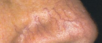
Blue nevus
The blue nevus is popularly called Mongolian; it is located inside the dermis and is not connected to the epidermis in any way. The blue color of the spot is due to the fact that nevocytes are located deep in the tissues. Such a spot can be located in the torso, face, hands and sacrum. The formation has a pronounced boundary; it has the shape of a regular circle. Mostly such moles are flat, but sometimes they can raise the area of skin where they are located above the rest of the skin. Such nevi are practically not susceptible to malignancy.
Causes of nevi
All pigmented nevi of the skin can be divided into congenital and acquired. This classification is not scientifically based, since it is still unknown when exactly clusters of melanocytes are formed. This division is based only on the time of appearance of nevi. If areas of the skin become dark in color in early childhood, then the formations are considered congenital. Acquired nevi appear in adolescents or adults.
The causes of congenital age spots are unknown. It is believed that pathological migration of melanoblasts occurs during intrauterine development. Risk factors include illness during pregnancy, medications or other chemical exposures, and hormonal imbalances. A possible cause of the appearance of congenital nevi is considered to be genetic predisposition. This explains the appearance of “Mongolian spots” in children of Asian origin.
Acquired nevi are considered more dangerous, since the frequency of transformation of these neoplasms into cancer is higher. However, according to other data, all areas of pigmentation are formed at the stage of fetal development, and their appearance indicates the adverse effects of provoking factors. Regardless of this, the following causes of nevi are identified:
- Hormonal changes in the body.
- Skin infections.
- Solar insolation.
- Visit to the solarium.
- Skin damage.
In fact, the identification of such provoking factors is quite justified. Hormonal imbalance develops during puberty and pregnancy. During these periods, the incidence of moles is much higher. In addition, age spots often appear in women using hormonal contraceptives.
Chronic skin pathologies (atopic dermatitis, acne) and physical damage often provoke the appearance of pigmented nevi on the face, neck and shoulder area. The main reason for the appearance of moles is considered to be solar insolation. Exposure to ultraviolet radiation not only affects the occurrence of pigmentation, but also increases the risk of malignancy of formations. Therefore, people with problem skin should not be exposed to direct sunlight for a long time.
Moles on the body - reasons for their appearance
To clarify the nature of the described skin defects, it is necessary to understand what a nevus is. It is an accumulation of immature pigment cells (melanoblasts) in the skin against the background of a violation of their migration. The exact reasons for this process are unknown, but there are suggestions why moles appear. Congenital factors:
- heredity;
- pregnancy pathologies;
- exposure to radiation, ionizing radiation during pregnancy;
- allergies, intoxication during fetal development;
- acute genitourinary infections of the expectant mother.
Purchased options:
- mechanical damage to the skin;
- hormonal surges;
- taking oral contraceptives;
- ultraviolet radiation;
- allergic diseases;
- dermatological infections and others.
Classification of pigment formations
Based on their histological structure, there are about 50 types of nevi. Of these, the most significant are 10 types of benign formations, differing in origin and clinical picture. Doctors distinguish 2 main groups of nevi, each of which includes several types of age spots. The classification is based on the risk of malignancy of the formation. The first group includes melanogenic nevi. The risk of malignancy of such formations is low. These include the following types of age spots:
- Papillomatous nevus. It differs in that it looks like melanoma: it has an uneven, bumpy surface and rises above the surface of the skin. The hair on this growth continues to grow, but changes color. Papillomatous nevus has a dark brown tint. The localization of such a mole is the torso, limbs and scalp.
- Mongolian spot. It has different shapes and large sizes. This congenital pigmented nevus occurs in most children of the Mongoloid race. It does not rise above the epidermis and disappears on its own by the age of 20.
- Halonevus refers to acquired benign skin formations. It has an oval or round shape. The peculiarity of this mole is that there is a light rim around it. Such a formation can be located on any part of the skin or mucous membranes. With age, the mole becomes lighter and disappears.
- Intradermal pigmented nevus. It is characterized by small size and peculiar localization: the neck area and skin folds. Appears most often during puberty. Under the influence of unfavorable factors, it can malignize into melanoma. A complex pigmented nevus has a similar structure. It is a small pigmented formation resembling a papule.
- Fibroepithelial nevus. This formation consists of connective tissue and rarely undergoes malignancy. It protrudes significantly above the surface of the skin and has a light brown or pinkish tint. This mole has a round shape and a smooth surface. The formation can be congenital, but more often occurs in older people.
Non-melanogenic nevi rarely become malignant, but they require observation. The lack of growth of such moles indicates a favorable prognosis.
Diagnosis and types of pathology
If you notice a sudden appearance of a large number of moles, consult a doctor immediately to prevent the development of skin cancer. Let's look at the different types of nevi, what they look like and whether they pose a threat to human health.
There are several types of pigmented neoplasms:
- dysplastic. This form of the disease occurs in adults and children and is characterized by uneven edges of the spot and a large diameter. It is worth taking a close look at this pathology, because in most cases it is from this type of nevus that melanoma is formed;
- blue. This type of nevus is acquired and affects lovely ladies who are over 30. Spots form on the thighs, abdomen, and less often on the face. In appearance, such formations resemble a small sphere protruding above the surface of the skin. Such a formation very rarely develops into a malignant tumor;
- borderline. This type is most often a congenital pathology, looks like a small tubercle, usually small in size, smooth to the touch; if you notice increased roughness, this is a reason to contact a specialist;
- giant. A very unpleasant sight, often this is a congenital defect; as the child grows, the formation grows, warts, cracks, and peeling appear. This form of nevus should be treated immediately, because such a formation is not only a cosmetic defect, which often develops into melanoma;
- Setton's nevus. Completely discolored skin forms around the spot;
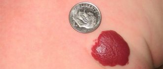
- "strawberry nevus" It is a red formation with vessels inside.
Depending on their size, nevi are divided into the following types: small (0.5–1.5 cm in diameter), medium (1.5–12 cm), large (12–20 cm), giant (more than 25 cm).
Diagnostics
Looking at the clinical picture, it will not be difficult for a specialist to identify a birthmark, but histological examination helps determine the type of nevus and the degree of threat to a person.
Only after conducting a series of tests will a dermatologist (or derma-oncologist) prescribe competent treatment. Using various ointments, creams, and tablets on your own is strictly prohibited.
Melanoma-dangerous skin nevi
Melanoma-dangerous nevi include benign skin neoplasms, the likelihood of which becomes malignant is high. Therefore, they require constant monitoring or radical treatment. Similar pigment spots include the following formations:
- Blue nevus of Jadassohn-Tiche. Despite the fact that it consists of differentiated pigment cells, the pathology refers to precancerous conditions. The blue nevus is small in size (up to 1 cm) and protrudes slightly above the surface of the epidermis. In some cases, it is represented by a nodule located deep in the skin. The formation has a purple or dark blue color.
- Borderline pigmented nevus. Refers to congenital formations. Such a mole protrudes above the surface of the skin and is dark in color. The color of the pathological area can be purple, brown or dark gray. The size of the nevus does not exceed 1.2 cm. The name of this formation is due to the fact that the pigment cells of which it consists are located on the border of the epidermis and dermis.
- Giant nevus. This pigment spot is large (more than 20 cm) and can occupy a significant area of the body. A giant nevus has a rough surface and dark color. Increased hair growth is observed in the pathological area of the skin.
- Nevus of Otta. A disease characterized by the presence of bluish spots on the skin of the face, lips, and mucous membranes of the eyes. It is often a congenital defect, but can also occur during adolescence. The risk of developing such a disease increases significantly among representatives of the Mongoloid race.
- Clark's nevus. It is characterized by asymmetry of contours, a flat surface and different colors. The size of the skin defect ranges from 5 mm to 6 cm. The nevus can be located in the back, along the back of the thighs, or around the genitals. It refers to dysplastic formations that are highly likely to transform into oncological pathology.
Melanoma nevi are a large group of pathological conditions that require diagnosis and treatment. In some cases, when removal of the formation is impossible, constant prevention of malignancy is necessary.
About pigmented nevus of the skin
A pigmented nevus (birthmark) is a benign skin neoplasm that occurs as a result of disruption of the normal structure of melanocytes—pigment cells containing melanin. They originate from the neural fold and, during embryogenesis, move to the basal (germ) layer of the skin of various parts of the human body. When the normal maturation or migration of these cells is disrupted, a congenital form of nevus occurs. Such deformed melanocytes (nevocytes) do not have processes, are characterized by poor metabolism and are capable of excessive pigmentation of the skin.
- Melanoform nevus was assigned an ICD 10 code – D 22.
What are pigmented nevi?
It can occur anywhere on the body and in any age group. It is most often found on the skin from birth, and increases in proportion to the child’s growth (in case of excessive growth, immediate consultation with an oncologist is required). One of the most common skin formations (80-90% of the population). Occurs more often in women. In representatives of dark races, pigmented nevi are less common than in whites. It does not cause discomfort to a person and has no symptoms even in case of degeneration (10-20%). For this reason, self-monitoring and periodic (once a year) examination by an oncologist are recommended.
In older people, pigmented nevi are more common due to age-related skin characteristics. Moles are extremely sensitive to external influences (direct sunlight, for example). They have a tendency to malignancy, especially in constantly injured areas. The only treatment option is surgical removal of the formation.
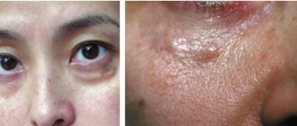
Nevus of the eye: features
The accumulation of melanocytes is observed not only on the skin, but also on the mucous membranes. An example is pigmented nevus of the eye. Another name for this formation is a benign choroidal tumor. It belongs to congenital pathologies, but begins to manifest itself only at 10-12 years of age. This is due to the fact that at this age there is an increased formation of pigment. There are 3 types of choroidal nevi:
- Stationary.
- Progressive.
- Atypical.
All of them belong to benign eye tumors, but under the influence of provoking factors they tend to malignancy. Like skin lesions, choroidal tumors are characterized by discoloration. So, what are pigmented nevi of the eye? Photos of such tumors are abundantly presented in the literature for ophthalmologists and oncologists, as well as on medical websites. Nevi are small spots in the eye, the color of which differs from the color of the iris.
Types of choroidal tumors
A stationary nevus of the eye is characterized by clear or feathery contours. It has a greenish or gray color. The shape, size and color of the formation do not change throughout life. Such tumors practically do not become malignant.
A progressing nevus is distinguished by the fact that it has a yellow rim around the main accumulation of pigment. The color and shape of the defect may change. In addition, such nevi increase in size, which increases the risk of vascular compression and a decrease in visual fields. Therefore, this type of pathology requires medical supervision.
Atypical nevi have a poor prognosis. Therefore, the slightest change or growth requires urgent surgical treatment. Such formations are light in color and are accompanied by deterioration of vision.
Diagnosis of pigmented tumors
If the nevus changes or appears, you should consult an oncologist. He will be able to make a differential diagnosis between various skin tumors and choose treatment tactics. An important study is dermatoscopy, which allows you to see the pigment spot under high magnification. If a tumor is suspected of malignancy, a complete laboratory diagnosis, chest x-ray and ultrasound of the abdominal cavity are performed. To establish the histological type of nevus, wide excision of the formation is performed. A biopsy is performed only in emergency situations, as it can lead to malignancy and spread of tumor cells.
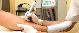
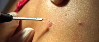
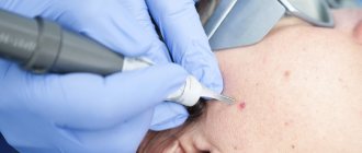
Pigmented nevus: treatment
Photos depicting nevi can be found in many sources on relevant topics. However, only a doctor can definitively determine what kind of tumor it is. Treatment of pigmented nevi is not always carried out. In cases where the mole is harmless and does not cause cosmetic discomfort, dynamic observation is indicated. Nevi, which are often subject to trauma, must be removed. You can remove the formation using liquid nitrogen or a laser. If there is a risk of malignancy, surgical removal of pigmented nevi is required. In this case, they retreat 2 cm from the spot and capture healthy tissue. The resulting material is sent to the histology laboratory.
When is treatment needed?
Currently, there are a variety of methods for treating and removing pigmented nevi. Among the main methods of therapy, experts primarily highlight the following:
- radio wave exposure;
- electrocoagulation;
- cryodestruction;
- laser exposure;
- surgery.
If any changes in the nevus occur, you should immediately seek help from a dermatologist or oncologist. This process may indicate the development of cancer.
If the diagnosis is confirmed, the doctor may prescribe other types of treatment. This applies to radiation, chemotherapy, removal of formations in various ways, etc.
A giant pigmented nevus is not dangerous in most cases. At the same time, it significantly affects a person’s appearance. In such situations, treatment of the disease is not indicated.
In order to remove the formation, plastic surgery methods are used. Their effectiveness directly depends on the location of the mole, its size, depth, type and many other factors.
Possible complications with nevi
The main complication of nevi is the transformation of normal pigment cells into melanoma. The following signs of malignancy are distinguished:
- Sudden increase in education.
- Bleeding or ulcers.
- Change in color of a mole.
- Painful sensations.
- Itching and burning.
If any of these symptoms are present, you should immediately contact an oncologist and have the tumor removed. Only timely surgical treatment will help avoid cancer.