Progress of the operation
Umbilicoplasty lasts from half an hour to one and a half hours. Before starting the procedure, the surgeon uses local anesthesia or general anesthesia. Further medical actions are based on the characteristics of the umbilical defect being eliminated:
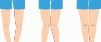
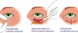
- The need for aesthetic correction. The surgeon makes an incision and forms the correct shape, indentation and parameters of the navel (usually a vertical classic fold);
- Elimination of excessive bulge. The doctor excises excess skin and inserts residual tissue into the skin fold;
- Recovery from loss. The navel is reconstructed from a flap of skin from the abdomen.
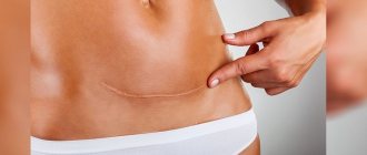
The specialist makes the incisions so that the scars after the intervention are located in the umbilical fold and are invisible to prying eyes.
Sometimes the navel is removed, but so far such an operation is not in great demand. It is mainly used by female patients who consider the navel to be an unaesthetic part of the body.
Buttock enlargement is no longer in fashion! Now everyone is doing navel surgery (6 photos)
Author: Eva Tushenkina
23 February 2020 15:10
Community: Facts
Tags: appearance plastic surgery plastic surgery plastic surgery plastic surgeon navel improvement
12369
6
According to data for 2020, Americans and American women spent $16 billion on improving their appearance through plastic surgery. Surgeons say that the fastest growing trend today is umbilical plastic surgery, or umbilicoplasty. Demand for it has doubled over the past few years! Patients ask for neat belly buttons, like those of famous actresses and models. A special excitement begins in the spring, when the fair sex thinks about a summer vacation.
0
Source:
See all photos in the gallery
According to plastic surgeon Matthew Shulman, the demand for navel surgery has increased thanks to social networks and the availability of any information. Another doctor, Dr. Thomas Sterry, believes that the navel is an important part of our anatomy from a cosmetic medicine point of view. And if you go to the beach and your belly button is not perfect, then you are unlikely to look sexy.
0
Source:
Dr. Shulman says the ideal belly button is a vertically oriented oval, which visually lengthens the belly and makes it appear flatter. “When it comes to belly button jobs, people bring pictures from Victoria Secret catalogs,” he says.
0
Source:
Also popular among Shulman’s patients are photographs of actress Jessica Simpson from 2005 (when she played in the film “The Dukes of Hazzard”) and model Emily Ratajkowski.
0
Source:
And sometimes they even ask for a “hidden” or “sewn up” navel. Yes, there is such a fashion now.
0
Source:
Very often, victims of umbilical piercing turn to surgeons. “I call it the Britney Spears effect,” says Dr. Shulman. “Those young girls who got their belly buttons pierced in the 90s are now grown, they are about 35 years old, and often their belly buttons look very sloppy due to weight gain or after childbirth.”
0
Source:
The operation is laparoscopic and is performed through small incisions made around the navel area. It is done under local anesthesia. Takes 30-40 minutes. According to Dr. Sterry, he performs at least 20 umbilicoplasties a year - and five years ago this figure was half that.
Source: - translated specifically for
Related links:
- The Iranian woman was eager to turn into Angelina Jolie
- The Twins Paid $1 Million to Look Like Brad Pitt, But They Didn't Work Out
- 63-year-old Kim Basinger scared fans with her frozen face after plastic surgery
- Japanese cosplayer spent $64,000 on a total makeover of her appearance
- Mother did not recognize her son after plastic surgery performed on him
subscribe to the “Facts” community
Tags: appearance plastic surgery plastic surgery plastic surgery plastic surgeon navel improvement
Did you like the post? Support Chips, click:
108 60 48
Liked
48 0
55
Partner news
Rehabilitation period
Hot bath
After the operation, for a couple of weeks it is necessary to follow medical recommendations to prevent suture dehiscence, swelling and hematomas in the intervention area:
- Refusal to take hot baths, replacing them with warm showers
- Protection of the surgical area from thermal effects (bath, stove heat, sauna)
- Regular massage, use of wound healing ointments and creams
- Refusal to lift weights and activities accompanied by tension in the abdominal muscles
For about a year after umbilicoplasty, it is necessary to protect the umbilical area from exposure to direct sunlight. During the first 2 months, specialists recommend wearing compression garments to reduce swelling and stimulate wound healing.
It is worth minimizing the consumption of foods that cause bloating and gas formation (plums, grapes, cabbage) - this can cause severe tension on the skin in the wound area.
The sutures are removed after about a week. The effectiveness of navel surgery is noticeable after about 1.5-2 months.
Umbilical hernia in adults
An umbilical hernia is a pathological condition in which abdominal organs protrude through the umbilical ring. Umbilical hernia in adults is not a common occurrence (about 5-12%) - it is more typical for newborns and infants. The main cause of this pathology is weakness of the anterior abdominal wall, which can develop due to various factors.
In this article, we will look at the main symptoms and main reasons why an umbilical hernia occurs in adults, what treatment methods exist, how the operation is performed, and also show what an umbilical hernia looks like in the photo.
Possible complications
- Infection. Can occur during surgery or due to poor tissue care. Accompanied by prolonged swelling, redness, and pain. Requires antibiotics before and after surgery;
- Rough scars. Associated with problematic healing or individual characteristics of the patient’s body. The problem can be corrected by using corticosteroids;
- Hernias. They appear when the depression is formed incorrectly (in this case, the tissues close to the problem area are injured). It is removed surgically.
Umbilical hernia in adults - reviews of the operation, symptoms and treatment
Methods of performing surgery to remove an umbilical hernia: laparoscopic, open, hernioplasty, laser removal. ⭐️⭐️ ⭐️ ⭐️ ⭐️ Duration of surgery, rehabilitation and possible complications. Forecasts and patient reviews
Read advice from our experts
Symptoms of an umbilical hernia are:
- compaction or protrusion in the navel area;
- expansion of the umbilical ring;
- pain in the middle of the abdomen during physical activity, coughing;
- nausea.
The disease can occur at any age in both men and women. But more often, umbilical hernia occurs in infants and people over 40 years of age.
Weakness of connective tissue and abdominal muscles predisposes to the appearance of a hernia in adults. Protrusion can occur with increased physical activity, heavy lifting, or chronic diseases accompanied by coughing. Excess weight, constipation, pregnancy, and ascites (accumulation of fluid in the abdominal cavity) also contribute to the formation of a hernia.
An umbilical hernia in infants can be congenital (due to improper formation of organs in the womb) or acquired. The latter occurs with frequent increases in pressure inside the peritoneum due to crying and slow healing of the umbilical ring. In some cases, infants' hernias are repaired non-surgically - with the help of massage.
The primary examination of a hernia consists of a visual examination of the patient by a general practitioner. Inspection and palpation are carried out in a horizontal and vertical position. If a hernia is detected, the doctor prescribes further examinations:
- Ultrasound of the abdominal organs;
- herniography – diagnosis using a contrast agent injected into the peritoneum;
- FGDS (for the study of a complex of diseases of the gastrointestinal tract);
- computed tomography.
To clarify the shape and size of the hernia, ultrasound diagnostics is sufficient. However, for a more detailed study and identification of concomitant diseases, additional methods are used.
There are a number of contraindications to surgery for umbilical hernia in adults and children. Therefore, it is necessary to carry out examinations in advance, the results of which may force the doctor to reschedule or cancel the intervention. Preparation before surgery to remove an umbilical hernia includes the following tests:
- ECG;
- general blood analysis;
- Analysis of urine;
- coagulogram;
- fluorography;
- tests for HIV, hepatitis, syphilis.
Contraindications to surgery to remove an umbilical hernia are:
- severe cardiovascular disease, recent heart attack or stroke;
- pregnancy, especially 2-3 trimester;
- old age – from 80 years and older;
- cirrhosis of the liver;
- renal failure;
- diabetes;
- varicose veins of the esophagus;
- infectious diseases in the acute stage;
- blood clotting disorder.
Hernioplasty is considered a simple operation, especially for small hernias. But even in emergency cases, when the hernial sac is strangulated or grows, surgical intervention, as a rule, goes well and is successful for the patient.
Removal of an umbilical hernia in adults and children can be performed using the traditional method (using tissue tension, without the use of an implant) or with the installation of an endoprosthesis . The implant is a mesh that strengthens the weakened part of the peritoneum and promotes its overgrowth with new tissue. Endoprostheses are made from different materials. Some of them dissolve over time, leaving behind their own tissues that have grown on top. Others remain in place and can be removed if necessary. Surgery using an endoprosthesis helps reduce the risk of relapse by strengthening the abdominal wall.
Laparoscopic method
About 25 years ago, it became possible to use a gentle method for removing an umbilical hernia - laparoscopic. Now this is the main and safest way to treat hernias. The operation is performed through small incisions, into one of which a camera is placed, and into the other - surgical instruments. Due to its low invasiveness, the risk of adhesions is reduced and the postoperative period is shortened. Sutures after laparoscopy are practically invisible. Laparoscopic surgery for umbilical hernia in women is performed during the intermenstrual period. With laparoscopy, both the tension method of closing the hernial orifice and the installation of a mesh implant are possible.
Single-port laparoscopy or the single-incision SILS method has been used relatively recently and has quickly gained the sympathy of both surgeons and patients. The disadvantage of this method is that the puncture hole is much larger than the standard ones.
Hernioplasty using Sapezhko, Lexer or Mayo methods
These are traditional methods of hernioplasty, which involve closing the hernial orifice using tissue tension. There are some differences in the course of operations and indications for them.
The Mayo method is more often used in overweight people. Two crescent-shaped incisions are made around the navel to capture excess fat deposits. Then the hernial sac is cut, adhesions and necrotic tissue are removed, and the internal organs are set into the peritoneum. The edges of the hernial sac are sutured horizontally. The aponeurosis (the so-called plate of connective tissue that gives strength to the abdominal wall) is sutured.
During surgery using the Sapezhko method, incisions are made in the form of vertical lines. After placing the organs in the abdominal cavity, layer-by-layer stitching of tissue is performed. Otherwise, the procedure is almost the same as the Mayo method.
The Lexer method is suitable for cases where the navel and hernial sac are inseparable. If necessary (large size of the hernia), the navel is removed during the operation. The doctor makes a crescent-shaped incision around the tumor. When the hernial sac fuses with the navel, it can be difficult to release its contents. The bottom or neck of the hernial sac is opened, and the organs are placed in the abdominal cavity. A suture is placed on the neck, and the sac itself is cut off. The hernial orifice is closed using the Lexer method by applying a silk suture to the aponeurosis around the ring and several sutures to the rectus abdominis muscles. After this, the skin flap cut out at the beginning of the operation is returned to its place and secured with interrupted sutures.
Removal of an umbilical hernia in adults, photo of an umbilical hernia
Removal of an umbilical hernia in adults is performed under local or epidural anesthesia.
Hernioplasty (hernia repair) can be performed either traditionally surgically or laparoscopically. Traditional removal of an umbilical hernia in adults involves making an incision in the skin and subcutaneous tissue. Then the hernia is either reduced inside the abdominal cavity (if the size of the defect is small), or it is completely removed. The next step is to strengthen the umbilical ring. For this, either the patient’s own tissues (tension hernioplasty) or synthetic materials (non-tension hernioplasty) are used. Removal of an umbilical hernia in adults using a non-tension method is more effective - it allows you to eliminate even large hernias and is characterized by a low likelihood of relapse. Recently, laparoscopic removal has become increasingly popular (photo above). It does not require an incision, is characterized by a short rehabilitation period, and a low risk of complications and relapses.
They will help to recognize such a pathology as an umbilical hernia in adults - photo. They can be found on various medical websites, as well as in patient reviews. The protrusion of the navel immediately catches the eye - in some patients it is more pronounced, in others less. The symptoms mentioned by the patient, palpation and ultrasound help the doctor confirm the diagnosis of a hernia. What does a strangulated umbilical hernia look like in adults? Photos will not help determine whether the hernia is strangulated or not. The differences between a strangulated hernia are symptoms such as acute pain, dyspeptic disorders, or disorders of intestinal function. Photos of umbilical hernia in men and women are above.