The main options for navel surgery
Have you been trying to heal your JOINTS for many years?
Head of the Institute for the Treatment of Joints: “You will be amazed at how easy it is to cure your joints by taking the product every day for 147 rubles .
Navel surgery is an aesthetic plastic surgery, most often it is performed:
Our readers successfully use Sustalaif to treat joints. Seeing how popular this product is, we decided to bring it to your attention. Read more here...
- Ladies after pregnancy and childbirth. Traditionally, within approximately 6 months after childbirth, the navel returns to normal along with the skin, muscle base of the abdomen and subcutaneous tissue. But, in some cases, the deformation of the navel turns out to be permanent; umbilicoplasty after childbirth can quickly eliminate the defect and restore the woman’s self-confidence.
- Patients after a bad piercing. The reasons for the deformation of the navel in this option will be: incorrect piercing technique, incorrect puncture of the navel ( or navel (Latin umbilicus) - a scar on the anterior abdominal wall remaining after removal of the umbilical cord in a newborn child
), violation of sterility, the body’s tendency to excess formation of connective tissue, allergy to iron objects. - Patients whose navel shape has changed after surgery. Deformation of the navel can be caused by surgical incisions in the area where the navel is located (along the middle strip of the tummy), tension of the skin of the tummy (plastic surgery to tighten the tummy without moving the navel).
- Men and women with acquired features of the umbilical ring structure. This is a bulging navel, formed due to improper handling of the umbilical cord in infancy or helplessness of connective tissues, which leads to the formation of an umbilical hernia and expansion of the umbilical ring.
- Older patients. Over the years, body tissues lose their elasticity, elasticity and sag downwards, age-related ptosis of the skin of the tummy leads to a change in the shape of the navel.
Main types of hernias
There are three main types ( VIEW: Literally: That which is visible to the eye
) hernias (lat. hernia): umbilical, inguinal and hernia of the snow-white stripe of the tummy.
The most common is a navel hernia .
It is also called fumes. In usual cases, when a child is born and there is no longer a need for umbilical cord vessels, the umbilical ring closes and is overgrown with connective tissue.
When the narrowing and closure of the umbilical ring does not occur, then an umbilical hernia develops, which is what we are talking about now.
In most cases, umbilical hernia in children goes away on its own.
An inguinal hernia never goes away on its own; it can only grow. Therefore, she must be operated on immediately. Urgently.
Hernia of the snow-white stripe of the tummy - what is it? We are talking about a ligament in the middle of the tummy. It is mostly small and only looks like a cosmetic flaw. If she doesn’t bother you, you can live with her. The operation consists of 2-3 stitches. In adults it only goes away with local anesthesia. These hernias themselves do not disappear. but they can be operated on when it is comfortable for you.
Why is surgery desirable? Because the bulge of the snow-white stripe of the tummy will spread even more during pregnancy in women and during sports exercises with lifting barbells or weights in boys. These are the individualities of the 3 types of hernial formations - in a nutshell.
Look at the photo of some other types of hernia:
What is an umbilical hernia and for what reasons does it occur in children?
The child, while in the mother's womb, is connected to her by the umbilical cord, through which he receives nutrients for formation and growth. After birth, the umbilical cord is tied up and cut off, and the umbilical cord is no longer needed.
Over time ( the form of physical and mental processes, the condition for the possibility of change
) the umbilical ring (
a round object with a hole inside (example: torus or solid torus)
) is tightened thanks to the muscles of the abdominal cavity. Since the umbilical ring in newborns is weak, from time to time it happens that it does not close completely, and this leads to the bulging of the digestive loop through it.
An umbilical hernia is a condition in which abdominal organs protrude under the skin through the umbilical ring. Most often, the disease is diagnosed in newborns, but it is also observed in one-year-old children and at 6-8 years old.
Umbilical hernia
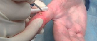
Girls, the surgeon diagnosed me with an umbilical hernia, surgery next week. I asked for laparoscopic surgery. But still, what does your navel look like after it, is there a scar, and how quickly do they let you go home? And another question everyone is asking is the drainage mesh? Oh, girls, I’ve written more than once, but still: maybe someone knows more gentle ways to remove an umbilical hernia and diastasis surgery? I looked at the photos of those girls who had it done in our city and by the same doctor, as I understood, he is the only one who does this in the city and was horrified - yes, the stomach becomes more neat, but they cut, cut how? There such scars remain, see under and so not neatly: compared to the scar on the cop and
Mode
But whether the patient is at home or in the hospital, he must be in bed for the first 2-3 days. That is, lie down all the time so that the stitches do not come apart due to an unusual overload for the body.
In some places, on the 3rd day, a person may begin to roll over and stand up. At the same time, it is necessary to realize that it is necessary to exclude any physical overload or fatigue for several days.
The next day after discharge and for a certain period (which is 7-10 days), the patient will have to visit the hospital for dressings. Later, the patient can perform the dressings himself. The nurse assigned to him will teach him this.
A week after the operation ( an action or a combination of actions to achieve a goal
) the patient is prescribed painkillers, medications and physiotherapeutic sessions that will promote rapid primary healing of the operated area.
In older people, after surgery, respiratory failure with tachycardia may occur. This may be a bad sign, which is best reported to the operating doctor.
When is surgery to remove an umbilical hernia prescribed?
This difficult disease is cured by a surgeon, and you need to contact him as soon as unpleasant feelings arise. Symptoms of an umbilical hernia include the following:
- pain in the abdomen when coughing or exercising;
- presence of nausea;
- extended umbilical ring.
An umbilical hernia can be diagnosed using several methods:
- Get examined by a specialist.
- Take an x-ray of the stomach and duodenum.
- Do an ultrasound.
- Undergo a gastroscopy function.
- To perform such a function as herniography is an x-ray method that involves introducing a special contrast agent into the abdominal cavity, which allows one to study the hernia.
Umbilical hernias can be of two types: congenital and acquired. Congenital can be found immediately after the birth of the baby. In the area of the navel, where the umbilical cord was, there is a spherical bulge with a wide base, running into the umbilical cord. If the baby screams loudly, the hernial bulge increases. How different congenital or acquired hernias can be can be seen in the video that is shown to clients in a medical institution. How to cure an umbilical hernia? Traditionally, surgical treatment of a hernia is not performed until the age of five. They are trying to eliminate it with the help of massage and healing physical exercise. If nothing helps and the navel does not miniaturize, you have to resort to surgical intervention for a hernia ( a disease in which internal organs protrude out of the cavity they normally occupy through a normally existing or pathologically formed hole in the
).
Types of operations
Surgical treatment can be performed either under local anesthesia, when only the intervention area is injected, or under general anesthesia. It is believed that in the first case the postoperative period is much easier. In the event that emergency surgery is performed, preference should be given to intratracheal anesthesia.
The patient is hospitalized on the day of the intervention. In total, you will have to stay in the hospital from three to five days. The manipulation itself lasts thirty minutes. If we consider other surgical diseases, then it is the hernia that is the easiest in terms of recovery. The operation can be performed in different ways.
In case of emergency surgery, preference is given to intratracheal anesthesia
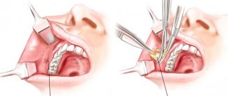
Olshausen method
This method is indicated in the presence of pathology in newborns. The incision is made directly on the hernial sac. The intestinal loops are reduced through it. If parts of the liver are also noted, then additional incisions are required. After this, all tissues must be sutured in layers.
Sapezhko's technique
The most common method used in modern surgery. After the onset of anesthesia, an incision is made in the immediate vicinity of the defect. Next, an incision is made near the neck of the sac. The organs are set through it, a suture is placed on the neck and the sac is cut off. After this, stitches are placed on the incision.
The recovery period after such manipulation is long and can take more than a year. Pain may be present for 2-3 months, which requires medication. Therefore, you should decide whether it is advisable to use this particular technique together with your doctor.
Sapezhko plastic surgery is successfully used to treat hernias of various sizes.
Not so long ago they began to use a technique that involves the use of implants. The technique differs in that in this case the tissues are not pulled together. A special mesh made of various materials is fixed at the defect site, which protects the weak spot.
It is especially beneficial to use the technique if there are large hernias, as well as if the defect has re-formed. The materials do not cause rejection and always take root easily. The technique has many advantages:
- rehabilitation after removal of an umbilical hernia is significantly reduced;
- the exit of the organs, that is, the umbilical ring, is reliably protected by a mesh;
- Such surgical treatment extremely rarely produces relapses.
The use of mesh implants accelerates the healing process of tissues after surgery
The technique is radically different from those described above. Laparoscopy of an umbilical hernia involves the creation of three punctures through which instruments are inserted. That is, there will be no rough scars on the stomach. If necessary, implants can be used.
At the same time, laparoscopic surgery may have some contraindications:
- dysfunction of the immune system;
- intestinal obstruction;
- circulatory disorders.
After surgery to remove an umbilical hernia, recovery is much easier using this method. In some cases, patients can return to work within a few days.
Intestinal obstruction is a direct contraindication for laparoscopic surgery
Umbilical hernia is a common pathology that occurs most often in newborns. The disease can also be acquired in nature, developing under the influence of reasons such as intense physical activity, heavy lifting, and diseases of the digestive system.
Why is an umbilical hernia dangerous? This question worries many patients faced with pathology. To better understand the possible health risks, you should find out what this disease is and whether an umbilical hernia is dangerous to health.
An umbilical hernia externally manifests itself as a formation in the form of a round or oval protrusion, in which the navel is not turned inward, but sticks out. The size of the formation increases with tension in the press; at rest, the hernia can be completely hidden.
In the early stages of development, the pathology manifests itself little; there are no signs of the disease. Less commonly, patients complain of pain, nausea, constipation and some other symptoms of the disease.
The reasons for the development of hernias are varied. Among the provoking factors are:
- Congenital pathology of the umbilical ring.
- Pregnancy and childbirth.
- Lack of physical activity.
- Intense sports activities.
- Lifting weights.
- Abdominal injuries.
- The patient is overweight.
- Rapid weight loss.
In the presence of provoking factors, the muscle gap through which the protrusion occurs increases, the formation reaches a larger size, the negative manifestations of the disease intensify, and the risk of complications arises.
Types of complications
An umbilical hernia is considered a ticking time bomb. The pathology may not bother the patient for years, but at a certain moment it can provoke serious consequences that are incompatible with life. The danger lies in the inability to predict the development of complications at various stages of the disease.
Organ inflammation
Incarceration of hernial contents
Depending on which organ gets into the hernial opening, its inflammation may develop. Most often, an umbilical hernia affects the omentum, stomach, and intestinal loops.
The complication is characterized by pain, general deterioration of health, swelling, redness of the skin, a sharp increase in temperature, nausea and some other manifestations.
The choice of treatment method depends on the degree of damage to the inflamed organ. In the majority of cases, surgical treatment is performed.
Fecal impaction
All hernias consist of a hernial orifice (an opening in the ligaments through which parts of organs protrude), a hernial sac and contents. With the development of pathology, loops of the large intestine can get into the hernial orifice. The process is accompanied by the accumulation of feces in the pinched areas and provokes intestinal obstruction.
Symptoms of this type of complication are:
- acute, persistent pain that subsides only after taking an anesthetic;
- increased body temperature;
- lack of stool;
- nausea, vomiting;
- flatulence, belching, prolonged hiccups.
Treatment of intestinal obstruction should be carried out as soon as possible, since the disease can cause severe abnormalities in the gastrointestinal tract.
This type of complication is considered the most common. Elastic strangulation occurs due to a loop of intestine getting into the hernial orifice, from where the organ cannot return to its place.
It is impossible to repair the hernia on your own. The patient experiences severe pain and deterioration in general condition.
The causes of infringement can be physical activity, prolonged exposure to an uncomfortable position, or a sharp turn.
Infringement can be recognized by the following manifestations:
- sharp pain;
- pulsation and sensation of movement in the affected area;
- deterioration of general health, weakness, irritability, sleep disturbance;
- bloating, heartburn, belching, nausea, vomiting;
- increase in body temperature to high levels;
- loss of appetite;
- the protrusion takes on a dense shape and does not move back into place.
If such symptoms develop, it is extremely important not to delay visiting a doctor. In complicated forms of infringement, serious disorders of the digestive organs develop, and the level of vitamins and minerals drops sharply. Less commonly, patients are diagnosed with problems with the cardiovascular system, breathing problems, and increased blood pressure.
The only method of treating various complications of an umbilical hernia is qualified medical care. Sometimes it is not immediately possible to transport the patient to the hospital. Before the ambulance arrives, it is recommended to adhere to the following rules:
- Provide a person with a state of peace.
- Apply a cool compress to the affected area.
- You shouldn't eat. If you are thirsty, you only need to take a few sips of water.
- In case of severe pain, you can take No-Shpu.
- Do not try to repair a hernia yourself.
Following simple recommendations will help support the patient and eliminate even greater complications.
When a person is admitted to the hospital, the diseased area is palpated. A doctor can determine the disease by visual examination, but the data obtained is not enough to make an accurate diagnosis. The presence of complications can be confirmed using special equipment. This:
- Radiography.
- Ultrasonography.
- Blood and urine tests.
Ultrasound examination of an umbilical hernia
If infringement and other complications are confirmed, emergency therapy is carried out, which in most cases consists of surgical treatment.
Indications for surgical intervention are prolonged pinching of parts of organs, intestinal obstruction, and the development of a severe inflammatory process. If the acute course of the disease lasts less than 2 hours, the pain subsides, the patient’s well-being improves - the pathology is re-diagnosed.
Treatment with surgery involves repositioning the affected organ to its anatomical location, suturing the hernial orifice through a surgical incision or punctures in the navel area. Recently, laparoscopy has been widely used. The method involves introducing video equipment into the peritoneum to the diseased area through small punctures.
During the operation, the surgeon not only puts the contents of the sac back into place, but also assesses the degree of damage to the internal organs, finds out whether there are bleeding and purulent tissue lesions.
Elimination of diastasis and umbilical hernia (PHOTO AFTER OPERATION)
I finally found a moment to write about my experience in eliminating diastasis and hernia after childbirth. In my case, due to the presence of a hernia, which was rapidly growing, only surgery was indicated; it CANNOT be removed with any exercises. Moreover, it is fraught with strangulation of the hernia, so the first thing I want to say is if the same trouble happens to you after giving birth, SEE A DOCTOR! to a competent surgeon! Unless the muscle diastasis is large, a course of special exercises will help; if there is also a hernia, such exercises are contraindicated in most cases.
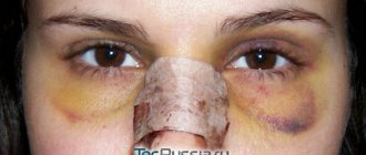
So. In order. My son is now one year and 3 months old, the pregnancy was difficult, polyhydramnios due to a large child, natural birth, the belly was huge during pregnancy. HUGE. I’ve always been tall and thin, my abs are almost perfect... I gained a little during pregnancy, but already in the 2nd trimester I realized that my stomach... to put it mildly... in short, I have to work a lot on myself after giving birth. And so it happened - the skin was flabby, covered in small light stretch marks, there was a 2-finger separation of the muscles (lucky, not too big!), and a hernia that was growing... at the time of the operation it was like half a quail egg and began to hurt, pull... when my son turned for a little over a year - I went to the surgeon - the verdict was ONLY OPERATION, it is very dangerous to walk with a hernia, especially since it gave me discomfort and there was a risk of strangulation (despite its small size). It is necessary to install a mesh and sew up the diastasis. I had it done at the clinic named after. Pirogov from St. Petersburg, this pleasure cost 100,000, doctor... the doctor is simply BRAVO. I installed a large enough mesh WITHOUT a vertical incision and a scar. Through the navel. When I looked at photos of similar operations, almost everywhere there was a vertical incision from the navel ((((I was very afraid of this! I don’t want to feel complex later, after all, you can’t disguise a vertical scar and almost always it is visible due to the presence of loose skin, folds form (But I was lucky) On March 31st I had an operation (4 hours, general anesthesia), on April 3rd I was already discharged. About the sensations after the operation... it was just a nightmare on the first day, on the second I was already up on my own. I spent a week at home almost constantly, it was pretty bad . I think the reason is still in the long general anesthesia. To do it beautifully, the doctors had to increase the operation time to 4 hours. After 2 weeks I was already running around the house after the baby))) But it was very difficult - you couldn’t pick him up! Nothing heavier 2 Do not lift kg at all for 2 months and MUST wear a special tight bandage. I still wear it during the day with breaks at night, although in theory it can be removed after 2 months. When pressing on the navel, there is now still a slight discomfort in one place with sudden movements I feel the mesh, BUT ALL THESE INCONVENIENCES ARE WORTH IT. The stitches were removed on the 10th day painlessly, there were only a few of them, only in the navel area. Now there is no fear of strangulating a hernia, and you can see the cosmetic effect in the photo! My waist has lost more than 4 centimeters, despite the fact that I have gained 3 kg after the operation)))) The navel is not quite perfect yet - there are small folds, but this is scar tissue - it should smooth out. At least I hope that my tummy will be completely smooth soon! I wish good luck to everyone who has encountered this problem. Decide to have surgery, if it is really necessary, let everything go well))
Sutures after surgery: features
Removal of suture threads is scheduled approximately one week after surgery. This procedure causes discomfort and some pain at the suture site. After removing the threads, doctors recommend continuing to wear a bandage or a belt that replaces it for several days. Absolute restoration of muscle tone and all connecting damaged tissues occurs two months after surgery. After this, you can completely stop wearing the bandage and return to your normal rhythm and lifestyle. However, rehabilitation depends on the individual characteristics of the body, and wound healing can occur at different times. The healing time also depends on compliance with the doctor’s instructions, diet and other factors. With any load on the patient’s abdominal area, the stitches may come apart or the internal ligaments and muscles may tear. Therefore, it is recommended to wear the bandage constantly until the muscles are completely restored.
Umbilical hernia in adults - reviews of the operation, symptoms and treatment
Methods of performing surgery to remove an umbilical hernia: laparoscopic, open, hernioplasty, laser removal. ⭐️⭐️ ⭐️ ⭐️ ⭐️ Duration of surgery, rehabilitation and possible complications. Forecasts and patient reviews
Read advice from our experts
Symptoms of an umbilical hernia are:
- compaction or protrusion in the navel area;
- expansion of the umbilical ring;
- pain in the middle of the abdomen during physical activity, coughing;
- nausea.
The disease can occur at any age in both men and women. But more often, umbilical hernia occurs in infants and people over 40 years of age.
Weakness of connective tissue and abdominal muscles predisposes to the appearance of a hernia in adults. Protrusion can occur with increased physical activity, heavy lifting, or chronic diseases accompanied by coughing. Excess weight, constipation, pregnancy, and ascites (accumulation of fluid in the abdominal cavity) also contribute to the formation of a hernia.
An umbilical hernia in infants can be congenital (due to improper formation of organs in the womb) or acquired. The latter occurs with frequent increases in pressure inside the peritoneum due to crying and slow healing of the umbilical ring. In some cases, infants' hernias are repaired non-surgically - with the help of massage.
The primary examination of a hernia consists of a visual examination of the patient by a general practitioner. Inspection and palpation are carried out in a horizontal and vertical position. If a hernia is detected, the doctor prescribes further examinations:
- Ultrasound of the abdominal organs;
- herniography – diagnosis using a contrast agent injected into the peritoneum;
- FGDS (for the study of a complex of diseases of the gastrointestinal tract);
- computed tomography.
To clarify the shape and size of the hernia, ultrasound diagnostics is sufficient. However, for a more detailed study and identification of concomitant diseases, additional methods are used.
There are a number of contraindications to surgery for umbilical hernia in adults and children. Therefore, it is necessary to carry out examinations in advance, the results of which may force the doctor to reschedule or cancel the intervention. Preparation before surgery to remove an umbilical hernia includes the following tests:
- ECG;
- general blood analysis;
- Analysis of urine;
- coagulogram;
- fluorography;
- tests for HIV, hepatitis, syphilis.
Contraindications to surgery to remove an umbilical hernia are:
- severe cardiovascular disease, recent heart attack or stroke;
- pregnancy, especially 2-3 trimester;
- old age – from 80 years and older;
- cirrhosis of the liver;
- renal failure;
- diabetes;
- varicose veins of the esophagus;
- infectious diseases in the acute stage;
- blood clotting disorder.
Hernioplasty is considered a simple operation, especially for small hernias. But even in emergency cases, when the hernial sac is strangulated or grows, surgical intervention, as a rule, goes well and is successful for the patient.
Removal of an umbilical hernia in adults and children can be performed using the traditional method (using tissue tension, without the use of an implant) or with the installation of an endoprosthesis . The implant is a mesh that strengthens the weakened part of the peritoneum and promotes its overgrowth with new tissue. Endoprostheses are made from different materials. Some of them dissolve over time, leaving behind their own tissues that have grown on top. Others remain in place and can be removed if necessary. Surgery using an endoprosthesis helps reduce the risk of relapse by strengthening the abdominal wall.
Laparoscopic method
About 25 years ago, it became possible to use a gentle method for removing an umbilical hernia - laparoscopic. Now this is the main and safest way to treat hernias. The operation is performed through small incisions, into one of which a camera is placed, and into the other - surgical instruments. Due to its low invasiveness, the risk of adhesions is reduced and the postoperative period is shortened. Sutures after laparoscopy are practically invisible. Laparoscopic surgery for umbilical hernia in women is performed during the intermenstrual period. With laparoscopy, both the tension method of closing the hernial orifice and the installation of a mesh implant are possible.
Single-port laparoscopy or the single-incision SILS method has been used relatively recently and has quickly gained the sympathy of both surgeons and patients. The disadvantage of this method is that the puncture hole is much larger than the standard ones.
Hernioplasty using Sapezhko, Lexer or Mayo methods
These are traditional methods of hernioplasty, which involve closing the hernial orifice using tissue tension. There are some differences in the course of operations and indications for them.
The Mayo method is more often used in overweight people. Two crescent-shaped incisions are made around the navel to capture excess fat deposits. Then the hernial sac is cut, adhesions and necrotic tissue are removed, and the internal organs are set into the peritoneum. The edges of the hernial sac are sutured horizontally. The aponeurosis (the so-called plate of connective tissue that gives strength to the abdominal wall) is sutured.
During surgery using the Sapezhko method, incisions are made in the form of vertical lines. After placing the organs in the abdominal cavity, layer-by-layer stitching of tissue is performed. Otherwise, the procedure is almost the same as the Mayo method.
The Lexer method is suitable for cases where the navel and hernial sac are inseparable. If necessary (large size of the hernia), the navel is removed during the operation. The doctor makes a crescent-shaped incision around the tumor. When the hernial sac fuses with the navel, it can be difficult to release its contents. The bottom or neck of the hernial sac is opened, and the organs are placed in the abdominal cavity. A suture is placed on the neck, and the sac itself is cut off. The hernial orifice is closed using the Lexer method by applying a silk suture to the aponeurosis around the ring and several sutures to the rectus abdominis muscles. After this, the skin flap cut out at the beginning of the operation is returned to its place and secured with interrupted sutures.
Why does such a hernia occur?
Causes of umbilical hernia and its manifestations:
- Obesity or overweight
- Connective tissue weakness
- Weakness of the muscles of the anterior abdominal wall.
Factors influencing increased intra-abdominal pressure in children:
- Severe and constant crying (if newborn baby)
- Constipation
- Frequent cough.
Symptoms depend on the size of the umbilical hernia, the presence of complications and other causes. The main identifying characteristic of a hernia in the abdomen is the presence of a small protrusion in the navel area, which indicates the exit of the abdominal organs beyond its boundaries through the hernial orifice.
At first, the disease may not create inconvenience or cause pain. However, after some time the hernia will increase and make itself felt. Therefore, you should immediately seek professional medical help to remove an umbilical hernia. If the protrusion of the umbilical hernia is large, it can cause severe pain, constipation, nausea, vomiting, and intestinal obstruction.
- Train your abdominal muscles, constantly keeping them in good shape
- Control your weight with proper nutrition and exercise
- Women need to wear a brace during pregnancy
- You should avoid strenuous physical activity as much as possible.
The first question that arises in patients with a hernial protrusion is: “How to get rid of an umbilical hernia?” There may be several possible answers to this question. The most common way to get rid of this problem is surgery. However, it is worth noting that recently medicine has been developing especially intensively, so you can choose alternative options.
An umbilical hernia in adults can cause many complications. This is damage, infringement, inflammation of organs, coprostasis (accumulation of feces in the intestines).
Preoperative preparation includes examination of the patient’s blood and urine composition, analysis for the presence of infections (HIV, syphilis, hepatitis), blood biochemistry, chest x-ray, electrocardiogram, coagulogram. Preparation for surgery includes checking whether there are chronic foci of infection in the body, and if they are present, it is necessary to carry out sanitation - preventive treatment aimed at preventing the development of inflammatory complications after surgery.