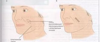
Langer's lines are conditional lines on the surface of the skin, indicating the direction of its maximum extensibility. They are named after the German anatomist, who in 1861 studied in detail the elastic properties of human skin (article “On the anatomy and physiology of the skin. On the splitting of the skin”).
description
They are round or ribbon-like, straight or crimped fibers of the connective tissue of the skin. If their number increases in a certain place, then they are connected to each other by branches in the form of a network, which easily stretches in the direction of the fibers, and then takes on its original appearance.
Langer's research also showed that the plexus of connective tissue fibers is a lattice-like formation of vascular bundles with loops elongated diagonally. The narrower the loops, the more parallel the vascular bundles are located. According to Langer, the direction of movement of the elastic fibers of the skin is constant and varies in different areas of the body.
The significance of Langer's lines in forensic medicine
Langer lines and skin strength
The strength properties of the skin depend on the direction of the acting force relative to the orientation of the collagen fibers (Langer's lines). The skin provides maximum resistance when the direction of impact coincides with the orientation of these fibers; the specific tensile strength of the skin along the Langer lines requires a load almost 3 times greater than in the transverse direction.
The magnitude of the maximum load leading to skin rupture
Changing the shape of damage
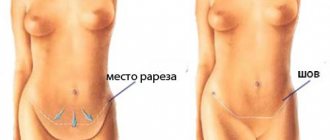
The shape of wounds on the skin, after removing the wounding object, changes its shape. For example, wounds from piercing objects with a ribless surface are not round, but slit-shaped, and their longitudinal dimensions in certain parts of the body are parallel.
There is no dependence of changes in the size of the skin preparation after exposure to fixing solutions relative to the location of Langer's lines.
Skin: functions and structure.
Now we will turn a little to biology, and consider the skin as a living organism. The artist is used to working on canvas, and since a makeup artist is a makeup artist, his main work surface is human skin, which in terms of scale is the largest organ, the area of which in an adult individual sometimes varies from 1.5 m² to 2. 3 m², and makes up approximately 15% of the total body weight. One of its main functions is protective: to protect the body from exposure to the external environment, free radicals, ultraviolet rays, and damage to internal organs. It provides external breathing, participates in thermoregulation, metabolic and excretory processes of the body. Like fabric - it is elastic, porous, durable, waterproof, antibacterial, sensitive. At low temperatures (cold), the lumens of the skin blood vessels are reduced, which means less heat transfer and more heat retention. When the temperature rises, the pores and blood vessels expand, then metabolic processes proceed faster and more intensely, sweating increases and the skin loses a lot of moisture. But do not forget that the intensity of heat transfer will also depend on the humidity of the air (climate), and the thickness of the subcutaneous tissue (including racial characteristics). We have looked at the main functions of the skin, now let's look at its structure. The skin consists of three layers: the outer - epidermis, the middle - the skin itself, or dermis, and the inner - subcutaneous fatty tissue. Each layer performs its own functions. The task of the makeup artist will be to work with the uppermost layer of the skin - the epidermis, since decorative cosmetics are designed for external use and should not affect the lower layers of the skin. Now we will touch in more detail on the structure of the epidermis and working with it, and only superficially then we will touch on the features of the functions and structure of other layers. The epidermis has a thickness of 0.07-2.5 mm. In appearance it resembles a narrow strip, although in fact it consists of 5 layers: horny, shiny, granular, spinous and basal (germ). The last layer contains such an important pigment as melanin. The upper layers of the epidermis become keratinized, forming the keratin (horny) layer, and in its lower basal (germ) layer, on the contrary, cells are constantly formed. Dead cells serve as a protective surface, but they constantly die and slough off, being replaced by new ones. The journey of a new cell from the basal layer to the keratin layer lasts approximately 2-4 weeks. In childhood, cell renewal occurs faster, due to the fact that the body grows; with age, this reproduction speed gradually fades away; visible aging processes begin to appear when the number of dead cells exceeds the number of new ones. Therefore, it is very important to take good care of your skin, cleanse, exfoliate, tone and moisturize it at least 2-3 times a day. In the morning, to put it in order after sleep and metabolic processes, because the body does not stop working when our consciousness turns off. For lunch, for those who have problem skin and well-developed subcutaneous tissue. This requires additional toning and fat removal. In the evening, since during the day it is exposed to external factors (sun, dust, environment), it sweats, removes metabolic products, thereby becoming polluted. Also, before applying decorative cosmetics to the skin, it must be cleaned of cells that have already been exfoliated during the day (with cleansing preparations or tonic), so that cosmetics can then be applied to clean, smooth and even skin. After all, the quality and cleanliness of the work will depend on the condition of the skin. There is even a saying: there is no better makeup than clean and well-groomed skin. Let's return to the epidermis: its deep layers contain pigment cells. They produce the pigment melanin, which affects hair color and skin color in this case, as well as the acquisition of one or another shade of tan. Melanins absorb ultraviolet rays, and thereby protect the tissues of the deep layers of the skin from radiation exposure, deactivate free radicals, and are a catalyst for many biochemical processes. Thus, it is an integral part of the body's immune system. For a makeup artist, the level of melanin determines the level of contrast a person has and their facial tone. The more melanin the skin contains, the darker it is. The largest amount of it is found in the skin of the Negroid race, Indians, Indonesians, etc. Most peoples who live in the equatorial and subequatorial zones, with equatorial and tropical climates, have a high percentage of melanin in their skin; as a result, dark and dark skin serves as protection from intense ultraviolet radiation. Slightly less melanin pigment is found in peoples living in the subtropical zone, with a Mediterranean and subtropical climate: Latin, Eastern, Asian peoples. The average melanin content can be conditionally attributed to people living in a continental climate. And the smallest percentage of pigment will be contained in the skin of people living in the subpolar and polar zones, i.e. among northern peoples, low pigment content also developed historically, due to low-intensity solar radiation, and simply the lack of need for protection from it. And due to the cold and reduction of the lumens of blood vessels, it will appear even lighter, bluer and thinner. The epidermis is practically impermeable to water and solutions based on it. Fat-soluble substances penetrate the epidermis better due to the fact that cell membranes contain a large amount of fat and these substances seem to “dissolve” in the cell membranes. Therefore, many preparations contain emulsifiers that “combine” the water and fat bases. There are also no blood vessels in the epidermis, and its nutrition occurs due to the diffusion of tissue fluid from the underlying layer of the dermis. The next layer of cells is the dermis, or skin itself. This is the inner layer of skin, which has a thickness of 0.5 to 5 mm. It consists of two layers: papillary and reticular. The reticular layer consists of loose connective tissue, which includes the extracellular matrix and cellular elements. The reticular layer consists of loose connective tissue, which includes the extracellular matrix and cellular elements. The basis of cells in the dermis is fibroplast, which synthesizes the extracellular matrix, including collagen, hyaluronic acid and elastin. The dermis contains hair follicles, a large number of blood and lymphatic vessels that provide nutrition to the skin, also participates in heat exchange, it contains pain and sensory nerves, as well as receptors (which branch into all layers of the skin and are responsible for its sensitivity). The excretory function in it is performed by sweat and sebaceous glands. The sebaceous glands secrete fat, which lubricates the hair and skin and makes it elastic, protects the skin from exposure to the external environment, makes the skin waterproof, bactericidal (sebum, together with sweat, creates an acidic environment on the surface of the skin, which has an adverse effect on microorganisms). Sweat glands participate in heat exchange, remove waste products such as water (in the form of sweat), thereby maintaining a constant body temperature, cooling it and preventing it from overheating.
Now we will look at the structural features of the extracellular matrix, which is synthesized by fibroblast, as well as its functions. The absorption of the extracellular matrix includes two main components: the fibrillar part and the matrix. The fibrillar part is the fibers of collagen, elastin, and reticulin that create the framework of the skin. Intertwined with each other, collagen fibers form a network that is located almost on the surface of the skin under the epidermis. This is the framework that gives the skin its strength. In the facial area, collagen fibers create a special dense network, which is strictly laid and ordered so that it forms lines of least stretch - Langer's lines, also known as massage lines. It is along these lines that massage is performed and cosmetics are applied so as not to stretch the skin and not provoke the formation of wrinkles (this massage is best performed with the weakest fingers - the ring fingers). At a young age, collagen fibers are quite strong and provide the skin with mobility and flexibility, while maintaining its elasticity and shape. All this can be compared to an armored bed, the base of which is a metal mesh. While the bed is new, the iron springs quickly return to their original position, but over time the springs begin to sag and the bed loses its shape. It’s the same with our skin - while we are young, our springs (collagen fibers) keep their shape perfectly, but with age they begin to sag. The matrix (matrix or amorphous component) in its structure most closely resembles a gel and consists of polysaccharides. The most famous of them are chitosan, seaweed polysaccharides, and hyaluronic acid. It is the components of the extracellular matrix, both amorphous and fibrillar, that create the skin from the inside. The saccharides themselves do not form fibers, but they fill all the spaces between the connective cells and fibers. It is through them that the interstitial transport of all substances occurs. As a result, it is the condition of the dermis (water content in the polysaccharide gel, the integrity of collagen fibers, etc.) that determines the condition of the epidermis and the healthy appearance of the skin.
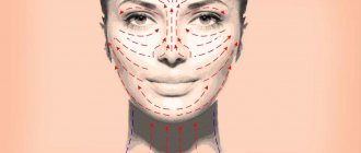
Drawing of massage lines
And the last thing we will touch on is the hypodermis or subcutaneous fatty tissue - the deepest layer of the skin. It consists of loose connective tissue, which contains many fat cells. The thickness of this layer varies and depends on lifestyle, nutrition, and metabolism. Fat is involved in thermoregulation and thermal insulation processes, preventing the body from overcooling or overheating, and also protects tissues and organs from mechanical influences. Fat cells are also depots in which fat-soluble vitamins (A, E, F, K) can be stored. Subcutaneous fatty tissue is very important as a mechanical support for the outer layers of the skin. Skin in which this layer is poorly expressed usually has more wrinkles and folds and ages faster.
Why is it so important to learn and always apply the Langer line rule?
Nothing preserves beauty and youth better than careful skin care and care from birth! Of course, every age has its own rules. The most important thing is not to miss the moment and prevent the premature appearance of wrinkles!
It would seem that everyone has heard about the existence of some mythical massage lines through which any cosmetic procedures must be performed. But, nevertheless, even those who know and have heard do not always follow this rule.
And in vain.
Of course, if you apply the same cream 1-2 times not along the massage lines, no disaster will happen. But the fact is that you apply the cream at least 2 times a day, which means 365*2=730 times during the year!
Therefore, you need to learn these lines, then integrate this skill into your life so that it becomes your useful habit!
To understand the very essence of this concept - the Langer line, I present to your attention a small encyclopedic reference:
“The lines of least stretch of the skin were discovered as a result of a study of the elastic properties of the skin by the German anatomist Langer in 1861. Therefore, experts call these lines Langer's lines.
The essence of this discovery is that collagen fibers in the skin do not lie randomly, but in a certain order. The stretchability of the skin depends on this - along the Langer lines the skin stretches 3 times less than across it. Wounds will form a smaller scar if the wound occurs along this line. Plastic surgeons and cosmetologists use this fact in their work.”
✅all movements of your hands on your face - whether you are applying cream, a mask, removing makeup or washing your face - should be done along massage lines☝
✅treat your skin as carefully as possible, because rough touches stretch the skin and make it sluggish
Science has known for quite some time about the existence of such a substance as collagen. These are protein threads present in connective tissue, namely in the intercellular substance. Collagen provides firmness and elasticity to fibers. This substance forms peculiar bundles. Langer lines are located in their direction.
History of discovery and subsequent research
In 1861, the scientist Langer published his work “On the anatomy and physiology of the skin. On the splitting of the skin." It was in it that he described the presence of conditionally drawn lines on the surface of the skin, along which it is most extensible.
Langer studied the properties of skin, such as elasticity, and noticed that skin is more stretchable in some directions than in others. He associated this phenomenon with the placement of collagen bundles under the skin in these places. He stated that in different places of the body, elastic fibers have different directions.
Along the Langer lines (cleavage lines) the strength of the skin is much higher. The scientist established this experimentally, using skin on corpses. In our time, researchers have tried to provide more accurate data about this phenomenon by doing experiments on animal skin. Of course, animal skin is significantly different from human skin. Therefore, the information obtained in this way left many questions.
Scientists at University College Dublin in Ireland and Professor Aisling Ni Annaidh personally tried to obtain accurate information. To conduct research, they needed about fifty-six skin fragments (taken from cadaveric material).
These studies confirmed Langer's hypotheses, but the question of the origin of this phenomenon remained open. This is probably simply a matter of the forces that act when attaching the skin to the body, but it could also be an anatomical phenomenon. That is, it is possible that the skin itself has hidden structures that form Langer's lines.
The main conclusion from all the studies of this phenomenon can be the fact that the existence of Langer lines simply needs to be taken into account by many specialists in different fields. From surgery to cosmetology, for a more effective effect on the skin you need to know the specifics of Langer's lines.
Langer lines on the face
To reduce the consequences after surgery, including plastic ones, as well as for more effective work of a cosmetologist, it is necessary to know where exactly the Langer lines are located on the face.
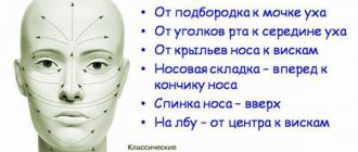
These lines on the face are located as follows:
- Along the line from the chin to the earlobes.
- From the corners of the lips to the middle of the ears.
- From the wings of the nose to the upper tips of the ears.
- From the middle of the nose to the temples.
- On the nose: from the tip of the nose to the bridge of the nose along the back and from the back of the nose to the wings.
- Upper eyelid: in a semicircle from the inner corner of the eyelid to the outer.
- Lower eyelid: in a semicircle from the outer corner of the eyelid to the inner.
- Forehead: from the middle of the forehead to the temples; from the eyebrow line vertically upward to the hairline.
- Neck: The front surface of the neck has an arrangement of fibers from bottom to top, while the side surface has fibers from top to bottom.
It is not so difficult to remember the location of these lines, but they should definitely be taken into account when cleansing your face and removing makeup, and even when applying make-up. These procedures should be carried out strictly in the direction of these lines, avoiding stretching the skin. Applying makeup to the surface of the nose is from the wings vertically to the base of the nose, and starting from the forehead, change the direction towards the temples.
The skin around the eyes is the thinnest and should never be stretched when applying or removing makeup. It is better to rub in the products with patting movements, and remove makeup with circular massage movements. Applying cosmetics to the face should occur along stretch lines, which will keep the skin more elastic. Proper care prevents the appearance of wrinkles.
Langer lines on the body and head
It is obvious that collagen bundles are located in a certain direction not only on the face, but throughout the body. For a better understanding, you should consider the drawing.
Langer's lines on the body are usually located in places where the skin naturally folds, since they are directed perpendicular to the muscles so that when the muscles are tense, the collagen bundles are not damaged. As we can see, Langer's lines are located horizontally on the hands, parallel to each other. Also in the center of the back and on the back of the legs. The lines run parallel to the ribs, bend around the pectoral muscles in front and the shoulder blades in the back. On the buttocks, directed from the center to the edges from bottom to top. On the front of the leg above the knee, the lines are located vertically. All these features are usually taken into account by surgeons during operations, massage therapists and cosmetologists.
When we need to determine the location of Langer's lines in places where there are no natural folds or wrinkles, we can do the following: squeeze an area of skin with your fingers, first along and then across. If skin folds appear, then Langer's lines are located there; if the surface is smooth, this area does not correspond to the direction of the lines.
Langer's lines are located not only on the face, but throughout the head. Their location is important to consider when undergoing hair transplantation, for example.
Langer's lines in the upper half of the forehead are parallel to the hairline.
The facial massage procedure is useful because it activates blood circulation and lymphatic drainage in the skin, which leads to an improvement in its condition. Also, when acting through the skin on the muscles through massage movements, you can remove the clamps in them, tone them, returning the natural youthful oval of the face. For a professional high-quality massage, a specialist must have certain qualifications and know where the facial massage lines are located.
In order not to stretch, but to tighten the skin, to give it elasticity during massage, applying cream and removing makeup, you need to know what the location of massage lines is on the face and neck.
Massage lines were discovered by the German scientist Karl Langer, who studied the structure of human skin and its elastic properties. “Langer lines” go in the direction of more elastic stretching of the skin.
Scheme of massage lines for the face and neck:
Location of massage lines:
- Line for neck massage, from the bottom of the neck along to the chin;
- From the middle of the chin to the earlobes in a smooth semicircular line, in both directions;
- From the middle of the top of the lip to the top of the ear in a smooth semicircular line;
- The nose is massaged from tip to bridge;
- Massage around the wings of the nose from the lip to the top of the wings;
- The cheeks are massaged from the nose to the upper tips of the ears;
- The lower eyelid is massaged from the outer corner of the eye to the inner, the upper eyelid is massaged from the inner corner to the outer;
- The forehead is massaged from the bridge of the nose with fan movements to the hairline.
Before the massage, a necessary condition is to clean the skin of makeup. The massage is performed with rich natural cream or oil.
HAIR.com
Langer lines
- lines of tension or layering of the skin that are characteristic of each part of the body. In microscopic sections made along these lines, most of the collagen bundles of the reticular layer are cut longitudinally, while in sections made across the lines we have cross sections of the bundles. The lines of delamination closely correspond to the lines of folds on the surface of the skin in most parts of the body. In other areas of the body, Langer's lines are visible or can be easily detected by squeezing the skin. On the scalp, Langer's lines are not visible due to the presence of hair and the large thickness of the skin. In hair restoration surgery, Langer's lines are often disrupted. The incision used for a sagittal elliptical reduction of the scalp along the midline (if it is not closed under tension) creates a cosmetically acceptable scar as it is oriented along Langer's lines (sagittally). As a rule, scars that cross these lines become wider after healing. The further the incision deviates from Langer's line, the wider the scar. Incisions made while lifting the scalp at the vertical temporal hairline and diagonally in the frontal region cross Langer's lines and always leave wider scars than those made sagittally along the midline. Incisions used posteriorly in Frechet triple flap slot correction are not parallel to Langer's lines and produce wider scars. These are just two examples of disruption of Langer's lines in scalp stretching procedures, and in my opinion the resulting scars are unacceptable.
A round dermatome or implanter (a punch-like tool) violates Langer's lines within 358 of 360 degrees (99.4%). Parallelism is maintained only within two degrees. An elliptical dermatome should result in smaller scars as there is less disruption of these lines. An incision made with a straight scalpel along the lines in the sagittal plane disrupts these lines to the least extent or may not violate them at all. Incisions 1.5 to 2 mm long, made with a microsurgical pointed scalpel and oriented along Langer's lines, do not leave scars if the size of the inserted graft (a flap of skin in the hair) is chosen correctly.
Hair restoration that can be performed with the least amount of scarring is the most aesthetically acceptable. Raised and recessed grafts, discoloration (hypo- and hyperpigmentation), and noticeable scars detract from the appearance of the skin and the perception of the result, regardless of the number of hairs transplanted per square centimeter. The scalp is the canvas on which we present our art, and the best results are obtained when the scalp undergoes the least amount of change.
How to do a facial massage
Before starting the massage, the skin on your face should be thoroughly cleaned and your hands should be washed thoroughly. The product you must choose is natural, without chemical ingredients.
When performing self-massage of the face, you should adhere to the rule of the four “Ps”:
- Punching;
- Pat;
- Tingling;
- Stroking.
All these movements should be carried out along the lines for facial massage with cream or oil applied to the skin.
- You need to start self-massage by pressing the skin with your fingertips;
- Then you should proceed to gentle rubbing in a circular motion in the direction of the massage lines;
- Next comes intense kneading of the skin and muscles underneath;
- Then pats;
- Finish the session with vibration and stroking.
Acupressure facial massage
During the procedure, acupressure may also be used. It consists of point pressure on strictly defined muscle areas. Such points are called active. Their stimulation helps to activate blood circulation, eliminate swelling, improve skin tone, which leads to the launch of a rejuvenation mechanism.
Here are some points whose regular massage will help in facial rejuvenation:
- in the center of the forehead (responsible for eliminating wrinkles in this area);
- on the inner and outer corners of the eyes;
- between the eyebrows in the bridge of the nose (light pressure on it can not only smooth out wrinkles on the forehead, but also generally improve well-being);
- in the center of the eyebrows, as well as along the entire eyebrow;
- on the temples (for headaches and lack of concentration);
- under the cheekbone lines;
- near the corners of the lips;
- under the nose and points near the wings of the nose;
- under the chin;
- under the lower lip (stimulation promotes relaxation and calmness).
Ideally, massage sessions should be performed daily. If this is not possible, then the option of a procedure every two to three days will also be good for improving the condition of the face.
Also on our website “Eco-Youth” you can read about the existence of...
Langer lines
1159. Vertical skin incision (semi-schematic).
1160. The main directions of tension of connective tissue fibers in the skin are the lines of tension and splitting of the skin, or “Langer’s lines” (left half), and points of irritation, or motor points (right half) (diagram).
( Anterior surface .) 1 - m. temporalis; 2—m. masseter; 3 - platysma; 4—m. sternocleidomastoideus; 5—m. omohyoideus; 6—m. sternohyoideus; 7—m. levator scapulae; 8—m. trapezius; 9 - m. sternothyroideus; 10 - m. deltoideus ( part starting from the clavicle ); 11—m. deltoideus ( part starting from the acromion ); 12 - m. pectoralis major; 13— m. coracobrachialis; 14—m. triceps brachii (caput longum); 15 - m. biceps brachii; 16—m. brachialis; 17— m. pronator teres; 18 - m. brachioradialis; 19—m. flexor carpi radialis; 20 - m. flexor digitorum superficialis; 21 - m. flexor pollicis longus; 22 - m. abductor pollicis longus; 23 - m. abductor pollicis brevis; 24—m. flexor pollicis brevis; 25 - m. opponens pollicis; 26 - m. abductor digiti minimi; 27—m. palmaris longus; 28 - m. obliquus externus abdominis; 29 - m. rectus abdominis; 30—m. tensor fasciae latae; 31 - m. pectineus; 32—m. sartorius; 33—m. adductor longus; 34 - m. rectus femoris; 36 - m. gracilis; 36 - m. vastus lateral is; 37 - m. adductor magnus; 38—m. vastus medial is: 39 - m. peroneus longus; 40 - m. tibialis anterior; 41 - m. extensor digitorum longus; 42—m. peroneus brevis; 43 - m. extensor hallucis longus; 44 - m. extensor digitorum brevis; 45 - mm. interossei dorsales; 46 - m. extensor hallucis brevis; 47 - m. abductor hallucis; 48 - m. soleus; 49 - m. gastrocnemius (caput mediale).
1161. The main directions of tension of connective tissue fibers in the skin are lines of tension and splitting of the skin or (“Langer’s lines” (left half), and points of irritation, or motor points (right half) (diagram).
( Posterior surface .) 1-m. infraspinatus; 2—m. teres minor; 3—m. deltoideus ( part starting from the spine of the scapula ); 4—m. deltoideus ( part starting from the acromion ); 5—m. teres major; 6—m. triceps brachii ( caput longum); 7—m. triceps brachii (caput laterale); 8—m. brachioradialis; 9—m. extensor carpi radialis longus; 10—m. anconeus; 11—m. supinator; 12—m. extensor carpi radialis brevis ; 13—m. extensor carpi ulnaris; 14—m. extensor digitorum communis; 15—m. abductor pollicis longus; 16—m. extensor pollicis brevis; 17—m. adductor pollicis; 18—mm. interossei dorsales; 19—m abductor digiti minimi; 20—m. extensor indicis; 21—m. extensor pollicis longus; 22—m. extensor digiti minimi proprius; 23—m. flexor digitorum profundus; 24—m. flexor carpi ulnaris; 25—m. triceps brachii (caput mediale); 26 - m. latissimus dorsi; 27—m. gluteus medius; 28 - m. gluteus maximus; 29—m. adductor magnus; 30 - m. biceps femoris (caput longum); 31 - m. semitendinosus; 32—m. biceps femoris (caput breve); 33—m. soleus; 34—m. flexor hallucis longus; 35 - m. abductor digiti minimi; 26 - m. flexor digitorum brevis; 37 - m. flexor digitorum longus; 38 - m. gastrocnemius; 39 - in. semimembranosus.
Incisions and suture lines on the face
Bold lines in the figures indicate incision lines, after which wounds can be closed with sutures without changing the direction of the line of these sutures.
The lines of force on the forehead run transversely; in the area of the glabella, in accordance with the direction of the folds, they bend between the eyebrows. The cuts should follow this direction of the field lines. On the upper and lower eyelids, incisions should be made parallel to the edge of the eyelid. If it is necessary to continue the incision of the upper eyelid, then the continuation line of the incision should be curved upward. Continuing the lower eyelid, the incision bends downwards at an angle of 60-80°. On the side of the face, incisions made parallel to the nasolabial fold should be considered correct. The cuts on the lips should be perpendicular to the border of the red border. In the chin-labial groove, the incision is made parallel to this groove, and on the chin itself - perpendicular to it.
Incision lines in the oral area: At the border of the oral area, incisions are made in the nasolabial fold or parallel to it. On the skin of the lips, incisions are made perpendicular to the border of the red border; they can be made at the border of the red border or parallel to this border, stepping back from it towards the skin by 1-2 mm.
Incisions on the side surface of the face and neck are made in the following directions: parallel to the edge of the scalp; at the base of the auricle—in the fold formed there or parallel to it; parallel to the angle of the lower jaw, if possible behind it. As you approach more central areas of the face, the incision lines should be more and more parallel to the nasolabial fold. Regular neck incisions run somewhat obliquely from a cranial-dorsal direction in a ventro-caudal direction.
On the back of the neck and on the back of the head, incisions usually run transversely.
If it is necessary to connect the incisions made on two eyelids, then this should be done so that a continuous semi-oval line of sutures does not form in the canthus, because in this case the scar shrinks, and as a result a scar “Mongolian fold” is formed. These cuts can only be connected in a Z-shape.
In the lower part of the upper eyelid, the incision is made parallel to the upper edge of the fibrocartilage, approximately 5 mm from it, from here it is directed upward and again follows parallel to the edge of the eyelid at a randomly chosen height within this area. Along the edge of the lower eyelid, an incision can be made directly next to the fibrocartilage, and in the area below it, so that it remains parallel to the palpebral fissure.
A longitudinal skin incision can be made on the nose in the area of the movable part of the nasal septum (pars mobilis septi nasi) or a transverse incision above the nasal openings. On the front of the neck, in accordance with the direction of the folds, the cuts should be directed transversely, slightly curving.
Picture: cicatrix optima pictures\119.jpg