Features of additional shares
Polymastia, or an additional lobe of the mammary gland, is included in the ICD-10 classification and refers to congenital developmental anomalies that are associated with deformities and chromosomal abnormalities.
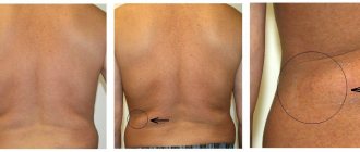
Polymastia is associated with an atypical position of the glandular tissue of the breast, most often indicating an additional lobe under the armpit.
Also, the breast lobule can be located between the mammary glands, directly under the collarbone or deep in the armpit. The most rare and remote cases of the position are outside the milky line, right between the shoulder blades or in the pelvic area.
Anatomical background
A lump-shaped swelling in the armpit area forms for a reason in this area. The localization of swelling in the cavity area is observed due to the large accumulation of lymph nodes in this anatomical zone, and the skin of the cavity is rich in sebaceous and sweat glands, as well as hair follicles.
The human body is designed in such a way that it always tries to limit and localize the inflammatory process. When an inflammatory process occurs in the armpit or upper limb, the axillary lymph nodes react first. In response to inflammation, they increase - this is one of the most common causes of the formation of swelling and infiltrates in the cavity.
It so happens that the armpit is a kind of depression that forms a fold of skin. In the armpit, the temperature of the outer skin is higher than in other places; it is also in this anatomical area that the most humid environment is observed, which, if there is insufficient hygiene in this area, leads to the development of an infectious process in it.
More than 90% of palpable formations in the axillary region belong to purulent-inflammatory diseases in a local form. In other cases, it is necessary to carry out differential diagnosis with more serious diseases.
Reasons for the formation of a false lobe
The appearance of an additional lobule of mammary gland under the arm is most often associated with genetic abnormalities and disorders during embryonic development. There are no other confirmed causes for this condition. However, doctors often identify factors that may contribute to the anomaly:
- severe stress of a pregnant woman, as well as poor diet and bad habits;
- genetic mutation at week 6, when the most important parameters of adipose tissue are established;
- sharp changes in hormonal levels during adolescence in a woman who has an additional lobe;
- hereditary predisposition - risks increase to 90% if the woman’s mother or grandmother had polymastia.
A separate factor that can be identified is that a pregnant woman takes certain groups of medications that contribute to the development of pathology: anticonvulsants, contraceptives, drugs to reduce the activity of the thyroid gland, as well as medications to normalize blood clotting. Chemotherapy, ionizing radiation and radiation treatments for cancerous tumors are especially dangerous.
Some household chemicals and industrial compounds with which a woman interacts can contribute to problems during pregnancy. Other factors cannot be ruled out:
- mental disorders of the expectant mother, frequent stress and emotional tension;
- chronic diseases;
- intrauterine infections;
- prolonged exposure to extremely low or high temperatures that do not correspond to the natural conditions to which a woman is accustomed;
- mechanical damage to the fetus in the 1st-3rd trimester after an accident, falls, blows.
Reasons for appearance
Polymastia is formed due to:
- Failures during embryogenesis as a result of changes in the genotype or its mutation. The formation of mammary gland lobules begins from the 6th week of fetal life. During this period, their number may exceed normal. At the 10th week of intrauterine development, their equalization occurs. In case of failures, as well as the negative impact of external factors, a violation occurs - the number of slices does not decrease.
- A sharp change in hormonal levels, for example in adolescence, as a result of a disruption in the synthesis of hormones or changes in their levels in the blood.
Hereditary predisposition to polymastia plays a significant role. Accessory mammary gland is more often diagnosed in representatives of the fairer sex who have had cases of polymastia in their family.
In the overwhelming majority, the cause of polymastia is a violation of embryonic development, which is associated with the following provoking factors:
- The mother's use of medications that have a detrimental effect on the fetus - medications, the use of which is not recommended during this period of gestation. Not recommended drugs include:
- anticonvulsants;
- medications whose action is aimed at normalizing blood clotting;
- drugs to suppress thyroid activity;
- oral contraceptives;
- chemotherapy drugs that are prescribed for oncology of various localizations.
- Poisoning with chemical compounds intended for use in domestic or industrial conditions.
- The harmful effects of ionizing radiation on the body of the expectant mother.
- The use of radiation therapy methods in the first weeks of pregnancy, as a rule, this happens when the woman’s position is not reliably known.
- Mechanical impact on the fetus during gestation, for example, abdominal trauma in the first trimester of pregnancy (fall from a height, road accident).
- Prolonged stay at very low or very high temperatures, which are considered abnormal in contrast to the woman’s usual living conditions.
- Bad habits of the expectant mother (smoking, drinking alcohol, narcotic and psychotropic drugs).
- Intrauterine infections.
- Psychological factors.
- Chronic diseases of the expectant mother, which worsen during pregnancy, leading to significant depletion of the body and disruption of compensatory mechanisms. This leads to disruptions during embryonic development, in particular during the formation of the mammary glands.
Symptoms of the development of polymastia
If the accessory mammary gland is small in size, it will not manifest itself for a long time. The following conditions can provoke symptoms of additional lobules:
- Pregnancy. The lobe becomes rounded and becomes distinct even before the delay occurs. Under the influence of prolactin, the mammary glands are transformed, which also affects the additional elements. This can be noticed by swelling, discomfort and even pain in the armpit area.
- Childbirth and lactation. On the 5th day, a woman experiences maximum milk production, which leads to breast enlargement, increased density and sensitivity. The same thing happens with the extra slice.
- The occurrence of inflammatory processes. Inflammation may or may not be associated with the feeding process. Often inflammation - mastitis - manifests itself long before and after lactation. Outwardly, it looks like an enlarged lymph node - the formation is quite dense, localized in the middle of the armpit. But inflammation can also indicate another disorder - a cancerous tumor. In this case, the formation may not hurt in the initial stages; it will be mobile, independent of other tissues.
- Development of mastopathy. In the axillary area, pain occurs in the second phase of the menstrual cycle, but with the onset of menstruation, the discomfort goes away. In some patients with mastopathy of the accessory lobe, a small compaction forms.
The additional lobe that appears does not have additional symptoms, such as fever or effects on general well-being.
What could it be
Do you have a lump under your arm and it hurts? Now let’s try to understand the options for the origin of this formation and why it is painful.
Most of these diseases are associated with the development of purulent-inflammatory lesions of the sebaceous or sweat glands of the skin in this area. Slightly less common are purulent-inflammatory diseases localized on the upper limb and causing lymphangitis and lymphadenitis, i.e. inflammation in the axillary lymph nodes.
- Hidradenitis Inflammation of the sweat gland, localized in the armpit. Popularly, hidradenitis is called a bitch udder, as it has a characteristic appearance. The sweat gland becomes clogged, its secretion accumulates in it along with bacteria located on the skin, then a local inflammatory process occurs, the gland enlarges in size and begins to rise above the general skin. The inflammatory process intensifies. Local edema occurs, hyperemia - redness of the skin in the area of inflammation, then due to the development of bacterial flora in this area, suppuration of the gland occurs. The appearance resembles a cow's udder, which is why it is popularly called so. Hidradenitis is a very painful inflammation, with which general manifestations of intoxication of the body are possible in the form of fever, general weakness, headache and other nonspecific symptoms of inflammation. In the case of hidradenitis, a red lump usually forms under the armpit.
- Lymphadenitis The second most common cause of lumps is lymphadenitis - inflammation of the lymph nodes localized in the armpit, while the lump under the arm hurts when pressed. The disease is the same as hidradenitis of a purulent-inflammatory nature. Lumps under the arm in women are a frequent manifestation of careless shaving of this area. As a result of microcracks and microtraumas caused by the razor, bacteria enter the thickness of the skin of the axillary area, which can lead to enlargement of the lymph nodes. Such formation can be either painful or painless - it all depends on the individual characteristics of the functioning of the body and the activity of the immune system.
- Furuncle This pathological condition is very similar to hidradenitis. However, in this case, inflammation of the sebaceous gland occurs. Blockage of the sebaceous gland can occur due to the peculiarities of the body’s hormonal system, for example, with diabetes mellitus or during puberty, as well as due to insufficient personal hygiene. The pathogenetic mechanism of the formation of the purulent-inflammatory process is practically is no different from hidradenitis, with the exception of the size of the inflammatory infiltrate. When a boil occurs, it is small in size, painful, with a local increase in skin temperature.
- Lipoma Popularly, a lipoma is often called a wen, and such a neoplasm occurs more often in young women. This formation is absolutely painless, easy to palpate and quite mobile. Lipoma occurs due to excessive development of adipose tissue in the area of the sebaceous gland, which leads to the formation of a kind of lump. In such a formation, a hereditary predisposition is clearly visible, and the course of the disease is characterized by slow growth and the absence of any other symptoms. Often, lumps under the armpits after childbirth occur in women with a hereditary predisposition, and the trigger in such a situation is a significant hormonal change in the female body in period of pregnancy and lactation. A lipoma or wen is purely aesthetic in nature and is not capable of causing harm to the body.
Health Hazard
If the appearance of an additional lobe of mammary gland under the arm is not associated with pain and unpleasant symptoms and it does not bother the woman in any way, there is no need to worry about the condition of the formation. In itself, it is not dangerous to health. The rate of growth into a malignant tumor is no greater than the formation of nodes in the normal mammary gland.
However, pathological processes in this part of the gland may be diagnosed too late, which will slow down treatment. It often turns out that it is possible to detect pathology even with an advanced abscess or stage 3-4 tumor. This can be avoided if you regularly undergo examinations by doctors - a gynecologist and a mammologist.
What does pain under the armpit in the chest indicate?
The doctor will first interview the woman in order to first understand what pain in the chest under the arm may indicate. First, the woman must tell how long the pain lasts, what methods can be used to relieve it, under what circumstances it occurred, and whether the pain radiates to other parts of the body.
Possible diseases that provoke the appearance of this symptom are:
- Muscle strains, injuries, falls from a height, blows to this area. Pain occurs immediately, which is why a person understands the reasons for its occurrence. After just a couple of days, if the shoulder is kept at rest, the pain goes away.
- After operation. Typically, operations on the chest or armpit area are accompanied by damage to the nerve fibers. Scars take a long time to heal, which is accompanied by pain. Women are often prescribed painkillers to relieve pain.
- Hidradenitis or lymphadenitis. We are talking about inflammatory diseases that develop in the sweat glands (hidradenitis) or lymph nodes (lymphadenitis). These diseases are accompanied by fever, acute pain in the affected area, pain in the arm and head. You should consult a doctor who will prescribe antibiotics and other treatment procedures.
- Mastalgia of a cyclical nature. These are pains that occur before menstruation in both breasts and go away on their own with their onset.
- Oncology. If a tumor is detected in the mammary gland, pain usually occurs in the later stages of the disease. Before this, the woman consults a doctor for help due to changes in the shape of the breast or nipple. The disease can be bilateral. The pain is first localized in the chest itself, and then moves to the armpit area.
- Disruption of internal organs. When the symptoms in question appear, we can talk about dysfunction of the cardiovascular or respiratory system.
- Osteochondrosis or neuralgia. Here you are encouraged to reconsider your diet and lifestyle.
go to top
Diagnosis of the condition of the accessory lobe
To make a diagnosis, doctors require precise instrumental examination techniques:
- Ultrasound. Very often, an additional lobe is found by chance during a comprehensive ultrasound.
- CT and MRI. They are used when a tumor is suspected and to clarify the nature of the tumor.
- Mammography. Allows targeted examination of the axillary and chest areas.
- Puncture. It is carried out for mastopathy and other signs of inflammatory processes to be sent for histological examination.
If there is a serious suspicion of cancer, a woman is prescribed blood tests to determine tumor markers.
It is impossible to diagnose an additional lobule of the mammary gland by visual examination and palpation alone. An experienced mammologist always prescribes an instrumental examination to accurately exclude unfavorable factors and pathological processes.
Need for surgery
Treatment of the additional lobe of the mammary gland under the arm is possible only by removing the pathological formation: in the presence of cysts, tumors, abscesses. In other cases, doctors do not recommend touching the lobule if it causes purely aesthetic discomfort.
Indications for surgery will be:
- detection of cysts, malignant tumors and fibroadenomas;
- regular pain and swelling of unknown origin;
- large size of the accessory gland;
- acute psychological discomfort.
Sometimes therapy with drugs for mastopathy is prescribed if the symptoms appear rarely and are mild. These medications are good at relieving swelling, discomfort, and mild pain. Hormonal contraceptives are often used to eliminate signs of polymastia.
Additional mammary glands under the armpit
If an ultrasound of the mammary glands revealed that there is an additional lobe under the armpit, do not be afraid. There is nothing dangerous about this. A little strange, but it does not pose any risk to life and health.
Additional mammary glands - developmental anomaly
During pregnancy and after childbirth, this anomaly becomes obvious. The additional lobe of the mammary gland, as well as the entire breast, receives milk, which can even drip from under the armpit or be released when pressure is applied to the duct.
Additional lobe of the mammary gland – what to do?
At the end of lactation, the additional glands can decrease to a completely invisible state and will not cause any inconvenience to their owner. But another option is also possible, when the lobules will still be visible, and when they become smaller, the skin under the armpits will sag. In this case, at the request of the woman, an additional lobe of the mammary gland is removed. The operation is performed under general anesthesia, and the recovery period lasts from 2 weeks to a month.
In general, doctors do not advise touching extra lobules if they do not interfere and do not spoil the appearance of the female body. Mammologists recommend simply observing additional glands and undergoing breast ultrasound more often.
Copying information is permitted only with a direct and indexed link to the source
https://womanadvice.ru/dopolnitelnye-molochnye-zhelezy-pod-myshkoy
Methods for removing an additional lobe
There are several methods for surgical removal of the accessory lobe of the mammary gland. The most common among them:
- Excision and suturing. Through an incision in the skin, the doctor removes the glandular tissue and the nipple, if present. The skin is then excised and the tissue is sutured. The removed tissue can be sent for biopsy to identify the causes of the formation of an additional lobule.
- Liposuction. During the operation, fat deposits are sucked out from the accessory lobule.
For up to 5 days after surgery, the doctor will install a drainage tube to prevent pus and other secretions from accumulating in the area. The recovery period takes on average 1 month.
Why does it hurt under my armpit?
There are a lot of reasons to talk about something specifically. Why does it hurt under the armpit?
- Received injuries to this area.
- Lifting weights.
- Perform operations in this area.
- Sprained muscles or ligaments of the shoulder joint.
- After sleep, when occupying an uncomfortable or incorrect position.
If a person has suffered a physical injury or muscle strain, he usually immediately begins to feel pain, which is why it is not difficult to diagnose the cause of its occurrence. It is much more difficult to understand that a dislocation or subluxation of the shoulder has occurred, since pain in this case does not occur immediately.
Also, various breast surgeries provoke pain, which is localized not only under the armpits, but also in the mammary glands themselves. During the operation, damage to the nerve fibers is quite possible, which begins to hurt several days after the operation.
If surgery was performed for breast augmentation, this factor should also be taken into account. Pain may occur under the arms due to the additional stress on the muscles and ligaments that now have to hold the chest. Along with the increase in breast size, weight also increased. The muscles and ligaments may not be ready for such weight, so at first they will hurt due to stretching and tension.
go to top
Prevention and prognosis
A woman who has this anomaly genetically cannot prevent the formation of an additional lobule. This is due to the fact that reliable information about the causes of the disease has not been collected. However, to avoid the growth of glandular breast tissue during pregnancy, a woman needs to stop taking provoking medications, reduce stress levels and monitor her health to exclude infectious and viral lesions.
If a woman has developed an additional lobe, she needs to be examined every year with an ultrasound. After 35 years, it is necessary to undergo mammography every 6 months in order to exclude cancer processes. It is not recommended to injure this area, as injuries can cause inflammation.
The prognosis for control of the additional lobule is favorable. The appearance of a malignant tumor in it is the exception in medical practices rather than the rule. It is also noted that after menopause, some anomalies may disappear without surgical intervention.
There are diseases that pose a threat to overall health. There are diseases and pathologies that require long-term treatment, but not urgent. And there are anomalies that affect the body, but do not require treatment, except for surgical intervention for cosmetic reasons. The mammary gland is the organ of the body that is most often subjected to surgery, removal of tissue, layers, tumors and lobules in the armpit.
Note that additional lobes can appear at different ages, behave differently and differ in the nature of growth and structure. Often, young mothers are horrified to discover swelling in themselves, run to surgeons in panic and ask to cut out an incomprehensible lump under the skin. It’s worth saying right away that there is no need to panic. Swelling can appear before childbirth, before pregnancy, during feeding, at any time. It is impossible to accurately predict the likelihood of a gland coming out in the armpit, since this is not a consequence of disease or pathology.
This is the most common possible anomaly, which in mammology is interpreted as an accessory gland. It appears only with any changes in the structure of the body structure inside the glandular tissue. More often there is a change in the genetic formation of the entire breast cavity. Under such circumstances, adolescents are susceptible to change. They have these neoplasms more often, but there is no need to worry. The lump in the armpit may hurt when menstruation occurs. After some time it does not cause any inconvenience, only embarrassment.
Some girls think and read that such a lobe may also have an additional nipple. No, this is excluded, since nipples can arise in the subclavian fossa without affecting the entire mammary gland.
Treatment methods and prevention
Women with deformed breasts very often undergo surgery. Surgical treatment is not mandatory.
This pathology does not interfere with feeding the child and can be asymptomatic. The operation is most often performed in cases of severe pain, severe cosmetic defects, functional disorders and a burdened hereditary history.
Postpartum surgery is not advisable as it may negatively affect milk production.
The additional lobe of the gland is removed by liposuction or removal of the formation, followed by suturing of the skin.
In case of a large formation, an incision of less than 1 cm is made during the operation. The adipose tissue is pumped out. It may be necessary to remove the glandular organ. In this case, the incision is enlarged.
The operation is performed under general (intravenous) anesthesia. Rehabilitation is fast. The sutures are removed after a week.
The axillary fossa heals quickly after surgery and does not cause discomfort. After treatment, you can lead a normal life.
If the operation is not performed, then dispensary observation is established for such women.
If you consult a doctor in a timely manner and undergo surgery, the prognosis is favorable. In some cases, complications develop.
This happens due to friction of the additional lobe on clothing or injury when washing the body. Possible damage during breastfeeding (BF).
There is no primary prevention of the anomaly, since it is congenital. In order to prevent complications, it is necessary to consult a doctor and undergo periodic examinations.
Thus, the presence of an accessory mammary gland in women does not cause physical discomfort.
The main method of treating patients is surgery. Conservative therapy is ineffective.
Polymastia is an abnormally located area of glandular tissue, similar to that which forms the mammary glands. Externally, the disease is a fully formed accessory gland of different sizes with an areola, nipple, and milk duct. Such an anomaly can be located:
- Between two normal mammary glands
- Below the collarbone
- In the armpit
- On the skin of the genital organs, near the anus, between the shoulder blades on the back (very rare)
Occasionally, an accessory gland is found that has only a nipple or only an areola, and even more rarely, false mammary glands with a nipple, which originate not from the glandular tissue, but from the adipose tissue. In most cases, pathology is detected in women.
The additional lobules of the mammary gland are somewhat different from polymastia. They do not have nipples, they look like a tubercle under the skin near normal mammary glands, but they behave like the latter (they are also susceptible to other diseases, and fill out during lactation). With age, the accessory lobules usually involute and may disappear completely.
The area of localization in the armpit is most likely for both polymastia and accessory lobules. Located in the armpit, the formation may be associated with the normal mammary gland, but not always. With this arrangement, the accessory gland is not only regularly injured, but also causes serious physical and aesthetic discomfort.
Symptoms of the disease
In any case, an extra lobe under the armpit is not normal. This is classified as a type of “disease” that can only be eliminated through surgery. But, like every disease, such growths cause symptoms in the body. Our body begins to react to abnormal phenomena occurring in the body. This is natural, so there are symptoms like any cold or flu. They are expressed as follows:
- The pathology is located in the left/right quadrant (top).
- There is swelling on the outside;
- There is a low temperature.
- No general malaise.
- Aching or dull weak pain.
- Breast asymmetry.
- The seal is movable, up to approximately 3 cm in diameter.
- A lump under the armpit hurts when menstruation begins.
Yes, these are not fevers and runny nose, by which you can determine the severity of the disease, call a doctor, and see all the control measures in a medical prescription. You can independently palpate and determine the severity of the tumor lobule. If a woman is breastfeeding, the tumor may not only hurt with each feeding, but also turn red and swell. Such an anomaly in the form of accumulations of glandular tissue cannot produce milk, and milk ducts are completely absent.
Medical diagnostics
The entire abnormal process of formation and development of the lobule under the arm cannot be diagnosed at the stage of development and formation. The presence or defect can be determined by the signs that a woman feels. To do this, the following procedures are additionally carried out:
- palpation by a mammologist;
- MRM (mammographic examination);
- Ultrasound;
- education puncture.
Mammography and ultrasound are needed in order to understand whether the lobe is a common defect in the development of the gland, or whether it is a benign tumor. Cancer in this case is impossible, since there are pronounced symptoms indicating the development of a viral formation.
With cancerous formations, fever and chills appear at the 3-4 stage of the formation of the disease. It is impossible to diagnose them, since symptoms appear long before the tumor in the glandular structure begins to develop and cover the surrounding tissues.
If you constantly injure the lobules, an inflammatory process will begin, which will lead to pain and mastitis of the additional lobe. It is difficult to recognize a disease in a place that should not be in the body, since the tumor-like lobe itself is a foreign body that needs to be punctured and removed. If such a lobe is found under the armpit of a woman over 40 years of age, additional testing may be necessary, such as an atypical test for cancer cells.
Treatment
To treat a lobule under the arm, surgery may be prescribed to remove it.
- The tissue is cut in the hole on the side of the formation.
- The lobules are excised and cut out.
- Cosmetic stitches are applied.
- Fluid from the empty canal is expected within 14 days.
If the glandular tissue and the lobe itself heal, then in the process lymph and ichor flow out from under the sutures. This is a natural process that occurs during any operation. After final healing, a woman is not recommended to plan a pregnancy for 6 months, so as not to complicate the formation of a healed wound after surgery. Otherwise, the woman may develop mastitis and bleeding.
In this case, you cannot take risks, even if the conception did not occur according to plan. It is recommended to terminate the pregnancy as early as possible, since in the second trimester the fetus, which is under the influence of hormonal levels, may suffer. He, in turn, has not yet been restored. If a woman has undergone surgery at the time of abrupt cessation of breastfeeding, there can be no talk of restoring lactation, even if the operation took 3 hours.
It is recommended to artificially maintain lactation in order to return to breastfeeding after 3 months. But doctors recommend stopping it completely so as not to cause complications for the mother and child. As practice shows, an additional mammary gland is easily removed without causing any negative consequences if you follow the regimen prescribed by doctors.
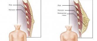
The mammary gland consists of approximately 10 - 12 lobes. All of them should normally be grouped together in the chest area. An accessory lobule of the mammary gland is an atypical location of part of the glandular tissue of the organ. A woman may not know about its presence for many years, since it is not always accompanied by any clinical signs. What are the dangers of an additional lobe of the mammary gland and does it need to be removed?
Read in this article
Forecast
The prognosis of extra breasts under the arm depends entirely on whether the woman is bothered by it or not. In any case, the anomaly should not be ignored. Any defect can lead to negative consequences.
If the patient does not want to resort to removal, then she must be constantly examined by a doctor. To do this, a visual examination and ultrasound are performed. This will allow you to control the growth and structure of the abnormal formation.
It is imperative to remove additional organs if they are susceptible to constant trauma. This occurs due to clothing, pressing by limbs, etc. Constant trauma can lead to a malignant neoplasm, which will entail a number of complications.
After everything is removed, it is quite possible to predict a favorable course. The occurrence of this anomaly cannot be called rare. It is important that the problem is correctly diagnosed and the degree of danger is adequately assessed. After which the issue of further treatment with surgical removal is decided.
Medical Expert Editor
Portnov Alexey Alexandrovich
Education: Kiev National Medical University. A.A. Bogomolets, specialty - "General Medicine"
The mammary gland consists of approximately 10 - 12 lobes. All of them should normally be grouped together in the chest area. An accessory lobule of the mammary gland is an atypical location of part of the glandular tissue of the organ. A woman may not know about its presence for many years, since it is not always accompanied by any clinical signs. What are the dangers of an additional lobe of the mammary gland and does it need to be removed?
Reasons for the appearance of an additional lobe of the mammary gland
It is necessary to distinguish between the concepts of “additional lobule” and “gland”. The pathologies are somewhat similar, but there are significant differences. The accessory breast (polymastia), in addition to glandular tissue, has a direct excretory duct, its own areolar zone and nipple. This is like a full-fledged mammary gland, only in a rudimentary size.
No reliable reasons for the formation of an additional lobe of the mammary gland have been established. The generally accepted opinion is that this occurs even during the period of embryonic development under the influence of some unfavorable factors. As a result, some of the cells remain in the armpit area, and all the rest migrate to the usual location of the breast.
This can be either the influence of harmful environmental factors, or medications in the early stages of pregnancy, infectious diseases during this period, etc.
A hereditary predisposition to this feature has been noted, so it is possible that the pathology is associated with mutations of some specific genes.
We recommend reading the article about lymph nodes in mastopathy. From it you will learn about the causes and symptoms of pathology, diagnostics and treatment methods.
And here is more information about lobular breast cancer.
Why does pain occur?
Only a doctor can say for sure why pain occurs under the armpit in the chest. A woman can only bear to speculate on what is happening to her. Here are some possible reasons to consider:
- Gynecological diseases.
- Changes in hormonal balance.
- Multiple abortions.
- Oncological diseases of the breast.
- Nervous strain and stress that constantly worries a woman.
- Hereditary predisposition.
It should be noted when the pain occurs. If it is periodic and is also localized in both mammary glands in the armpit area, then pay attention to the period of its occurrence. These may be the days before your period. Quite often, women experience such pain, which is completely natural. They are usually localized in the front of the mammary glands, but can also affect the armpit area. No treatment is required here.
Mastopathy becomes another cause of pain in the armpits. It is usually diagnosed in women between 35 and 55 years of age. This disease is characterized by neoplasms that are localized in one (less often in both) mammary glands. The disease becomes a consequence of hormonal imbalance, for which treatment is quite simple and effective.
go to top
Symptoms of an additional lobule under the arm
Small additional lobules may not show themselves for a long time. Typically, a woman becomes aware of their presence under the following circumstances:
- During pregnancy, sometimes even before a missed period. This is due to the fact that under the influence of prolactin, the mammary glands are transformed for subsequent lactation. Moreover, all these changes occur in the additional lobule. Swelling appears in the armpit area, possibly even discomfort or pain.
- After childbirth, especially if the girl is supporting lactation. On the 3rd - 5th day, the maximum milk supply normally occurs. At the same time, the breast increases significantly, becomes dense and sensitive to the touch. Everything proceeds similarly in the additional lobe.
Typically, 3–5 days after childbirth, women notice pain in the axillary region and upon palpation a tumor-like formation is detected. There is no need to do anything in such situations, everything will go away on its own in 2 - 3 days. In rare cases, if the outflow of secretions from the additional lobule is disrupted, inflammation may develop in it - lactostasis and mastitis, with an increase in temperature and a deterioration in the general condition. Treatment follows the general rules for this pathology.
- If an inflammatory process occurs in the accessory lobule or other diseases (even cancer). Lactostasis can occur at any time during breastfeeding, and non-lactation mastitis of the accessory lobules (inflammation at any age) also occurs. In this case, a woman may think that her lymph node has become inflamed, a boil has appeared, or something similar. With the same frequency as in a normal mammary gland, a cancerous tumor can arise in an additional lobule. Typically, a woman discovers a small formation in her armpit, painless, mobile or not relative to other tissues. Therefore, it is important to conduct research in this area during mammography or ultrasound.
- With the development of signs of mastopathy. The disease can occur at any age in different forms. The main symptoms are the appearance of soreness in the armpit area in the second phase of the cycle, everything goes away with the onset of menstruation. Sometimes even the appearance of a small compaction is detected, which regularly disappears and appears again.
So, you can suspect pathology if you have the following symptoms:
- the appearance of a compaction in the axillary region, less often it is the subclavian and others;
- formation is painful or not;
- Almost always the occurrence of symptoms is associated with pregnancy, childbirth, and the menstrual cycle.
Polymastia
- Between two normal mammary glands
- Below the collarbone
- In the armpit
- On the skin of the genital organs, near the anus, between the shoulder blades on the back (very rare)
Occasionally, an accessory gland is found that has only a nipple or only an areola, and even more rarely, false mammary glands with a nipple, which originate not from the glandular tissue, but from the adipose tissue. In most cases, pathology is detected in women.
The area of localization in the armpit is most likely for both polymastia and accessory lobules. Located in the armpit, the formation may be associated with the normal mammary gland, but not always. With this arrangement, the accessory gland is not only regularly injured, but also causes serious physical and aesthetic discomfort.
The development of the mammary glands starts from the 6th week of the embryonic period. The emerging strands of epithelial tissue grow, form ducts, and their excess parts disappear by the 4th month. If this process fails, a person may be left with an extra lobule or gland after birth.
Hormonal imbalances can lead to the formation of additional lobules even throughout life. So, in some women they were discovered only during pregnancy. Sometimes pathology is detected during puberty.
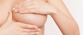
Signs and symptoms
Outwardly, it is almost impossible not to notice a problem in the body, so the patient’s psychological discomfort can be significant.
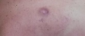
The lobules and glands protrude outward, sometimes resembling a normal mammary gland, but more often looking like a small lump on the skin.
Symptoms may include:
- Nipple soreness
- Aching pain in the gland itself
- Increase in size during menstruation
- Milk discharge from the nipple in nursing mothers
In some patients, oncological formations develop in additional lobules or with polymastia.
Diagnosis of the accessory lobule of the mammary gland
Pathology can be detected and confirmed using available methods. Namely:
| Method | Diagnostic features |
| Inspection and palpation | Can give general ideas about education. But if the lobule is undeveloped, there are no changes in it, it is impossible to identify it. |
| Ultrasound examination of the mammary glands | Often, an additional lobe is discovered by chance, especially if the woman is not bothered by it. |
| CT or MRI | They are used less frequently, for example, to clarify the nature of the formation or suspicion of a tumor. |
| Mammography | To identify it, it is necessary to specifically examine the axillary region (or subclavian, etc.). |
| Puncture | If necessary, especially in case of mastopathy, a puncture can be performed followed by a histological examination of the obtained material. |
The cause of pain is mastitis
Women often experience breast pain during breastfeeding. At the very beginning of lactation, such sensations become quite natural. The breasts are not yet accustomed to feeding, especially if a woman is giving birth for the first time. However, over time the pain should go away.
If the pain does not go away, then you should consult a doctor. Mastitis may be detected - a disease with the appearance of lumps due to stagnation of milk in the breast or irregular feeding of the baby. When the breasts are not cleared of milk at a certain frequency or the milk ducts do not completely empty the lobes in the mammary gland, stagnation begins. It leads to infection entering the area of stagnation through small cracks and wounds and provoking an inflammatory process.
At the initial stage, treatment will not require weaning the baby from the breast. The woman will continue to feed him as one of the ways of her recovery. However, at later stages, it may be recommended to stop feeding the baby for a short time so that the woman can be effectively treated.
Is education dangerous for health?
The extra lobe itself is not dangerous to a woman’s health. Many are afraid that it will eventually turn into a malignant tumor. In fact, the probability for this pathology is exactly the same as for all other women.
The difficulties are as follows. In some situations, pathological processes in this area may be diagnosed late due to a somewhat atypical clinical picture. As a result, you can get an advanced process, for example, with inflammation - an abscess, with a tumor - stage 3 - 4, etc.
Is it necessary to have surgery?
If the additional lobule of the mammary gland is not complicated by anything (abscess, cyst, tumor, etc.), there is no need to remove it. Even if it brings tolerable discomfort during breastfeeding or on the eve of menstruation, this is not an indication for surgery.
Removal of the additional lobe is recommended in the following situations:
- with large sizes;
- if it is accompanied by severe pain, swelling;
- if a cyst, fibroadenoma or even a malignant tumor is found in it;
- at the request of the woman, if it brings her psychological or cosmetic discomfort.
If a girl is bothered by some minimal symptoms, you can try treatment with drugs intended for the treatment of mastopathy. This way you can reduce the manifestation of pain, discomfort, swelling, and slightly reduce the size of the formation. Using properly selected hormonal contraception also helps relieve all symptoms.
Watch the video about benign breast tumors:
Removing the extra milk lobule
Operations to remove additional lobules of the mammary glands can proceed in different ways. It all depends on the size of the education, the desire of the woman, her age, whether she is going to give birth, etc. The approach is individual in each case.
Similar operations are performed by both oncologists and plastic surgeons. It is better to give preference to the latter if the lobule is not removed due to a tumor or abscess, since these doctors in most cases have more experience in correcting “extra” areas of skin, etc.
How is the operation performed?
With age, glandular tissue both in the mammary gland and in the accessory lobule is replaced by adipose tissue. This influences which surgery should be preferred. If, based on the results of the examination, it is determined that most of the lobule is adipose tissue, conventional liposuction can be performed. To do this you will need to make one small incision. Upon completion of the operation, it sometimes becomes necessary to install drainage to drain fluid for up to 3 to 5 days.
If the operation is performed at a young age, glandular tissue most likely predominates in the additional lobule. In this case, the entire gland should be removed. The steps of the operation are approximately as follows:
- Tissue incision over the accessory lobule or other cosmetic access.
- Removal of glandular structures and fatty tissue.
- If necessary, tissue plastic surgery, including removal of excess skin flap.
- Layer-by-layer suturing of tissues, if necessary, installation of drainages for the outflow of pathological fluid.
Recovery after deletion
The recovery period depends on the volume and type of surgery and ranges from a week to a month. For some time after the intervention, the woman may feel discomfort in this area. The main recommendations for preventing complications after surgery are as follows:
- It is necessary to follow all doctor's recommendations for treating the wound and taking medications. This will help prevent the wound from becoming infected.
- It is advisable not to become pregnant for 6 to 12 months, otherwise it may cause a relapse, especially if it was technically difficult to remove all the glandular tissue.