9801
0
A malignant tumor that is localized in the basal layer of the skin and spreads to healthy skin is called melanoma. Its development is ensured by pigment cells that have degenerated into cancer cells.
This disease appears due to the degeneration of an ordinary and harmless (at first glance) nevus or mole on various parts of the human body or in the mucous membrane. It proceeds aggressively and quickly. Melanoma affects both men and women aged 35 to 50 years.
Doctors are not always able to diagnose this disease at an early stage. It is detected when metastasis appears. This is why it is difficult, and in some cases impossible, to cure melanoma.
What it is
Melanosis, or melasma, is the formation of coffee-colored pigment spots on the skin.
Pathology most often occurs in women against the background of hormonal imbalance, when the concentration of progesterone and estrogen in the body does not correspond to the norm.
Pigment spots form in women:
- carrying a child;
- using oral contraceptives;
- resorting to hormone therapy during menopause.
Pigment spots are formed under the influence of sunlight. They occur in women living in southern countries. Summer is a period of risk. Increased solar activity provokes the development of pigmentation on epithelial tissues.
Causes
Doctors have identified several factors that cause melanosis of the skin. The causes of pathology include:
- concomitant diseases affecting the pituitary gland, ovaries and adrenal glands;
- impaired functioning of the thyroid gland;
- intoxication of the body with toxic substances - arsenic, resins and other aggressive hydrocarbons;
- infectious diseases prone to progression - syphilis, tuberculosis, dysentery, malaria;
- pediculosis in severe form with damage to the mucous membranes;
- avitaminosis;
- poor nutrition, leading to disruption of metabolic processes in epithelial cells;
- heredity (pathology is transmitted mainly from mother to child).
The causes of melasma are interconnected with the type of pigmentation, concomitant diseases and genetic predisposition to certain pathologies. When drawing up a treatment regimen, the doctor selects drugs that can combat the cosmetic defect and the root causes of the disease.
Types of melanosis
Doctors distinguish between several types of pathological pigmentation. The classification of the disease is based on provoking factors and symptoms. There are 5 types of melasma:
- Uremic. Occurs against the background of renal failure.
- Hepatic. Caused by liver disease (usually cirrhosis).
- Kakhetic. Caused by severe forms of tuberculosis, impaired functioning of the adrenal glands.
- Endocrine. Caused by diseases of the thyroid gland, a disorder of the pituitary gland.
- Toxic (arsenic). Appears against the background of severe intoxication caused by arsenic and hydrocarbons. Gasoline, kerosene, motor oil and other petroleum products lead to poisoning.
In addition to the general large categories, specific types of melanosis are distinguished. These diseases are severe and threaten the health and life of patients. Such pathologies are eliminated mainly by surgical methods.
The following types of melasma are distinguished:
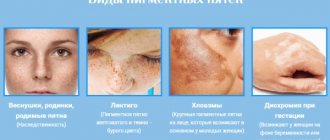
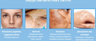
- Chloasma. The development of pathology is provoked by hormonal imbalance. Occurs in women during pregnancy and menopause.
- Becker's melanosis. Benign spots appear in young men.
- Dubreuil's melanosis. The disease affects women over 50 years of age. The formations can degenerate into cancerous tumors.
- Lentigo. Flat pigmented nevi appear on the epithelium.
- Moynahan syndrome. A huge number of brown spots that are benign in nature form on the epidermis of patients.
Stages
Depending on the degree of progression of the disease, the damaging ability of atypical formations and the patient’s health status, four stages of the disease are distinguished:
- Stage 1 – initial. Abnormal cells are just beginning to develop. Only the superficial epidermal layer of the face is affected. The tumor is immobile and can be easily treated if the anomaly is detected in a timely manner;
- 2 – stage of transition to deeper layers of soft tissues of the face . The size of the pathology is about 5 mm, with germination into internal tissues of 1.5 - 2 mm. Metastasis is almost always absent, the compaction does not leave the primary localization. At this stage, minor pain may appear;
- 3 – stage of active spread and penetration of cancer cells into lymphatic tissue. Accompanied by ulcers and increased body temperature;
- 4 – the final stage of the disease . The stage is severe and symptomatic, treatment is ineffective, all that can be done is to help the patient endure the manifestations of the disease and, as far as the situation allows, to extend his life threshold. At the moment, the liver and lungs are already affected, and the general toxicity of the body has reached a critical point.
Symptoms
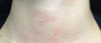
Pigmentation on the epithelial tissues of the face is the main symptom of melanosis. Brown-brown defects cause incredible psychological and physical anxiety. Unnatural coloring affects the skin of the forehead, cheeks, nose, and upper lip. The spots are located symmetrically, appearing simultaneously on both sides of the face, armpits, genitals and other places on the body.
Common symptoms of the disease include:
- diffuse pigmentation of epithelial tissues (found in newborns);
- hypersensitivity of the skin to ultraviolet radiation;
- merging of affected areas into large foci;
- swelling;
- compaction and coarsening of the stratum corneum;
- atrophy of epithelial tissues;
- impaired functioning of the nervous and endocrine systems.
The affected skin is constantly irritated. Patients feel itching and burning in the area of age spots.
Treatment
The optimal option for therapeutic treatment of cancer is selected individually by the oncologist, taking into account how far the process of organ damage has progressed and what form of the disease the patient suffers from. Based on these criteria, apply:
- removal is the most common way to solve the problem. Not only the tumor neoplasm is amputated, but also the lymph node connections in the case of metastases. At the same time, healthy surrounding tissue fragments are also partially removed;
- laser coagulation - is carried out when the anomaly is not too large, and the degree of its growth into the deep layers is insignificant. At the same time, the volume of capture of healthy areas is about 1 cm;
- photodynamic therapy - during the manipulation process, the irradiating effect on the pathology is achieved as a result of the introduction of photosimbelizers to the site of the lesion;
- Chemotherapy is carried out mainly in a comprehensive manner. Autonomous use of cytostatic drugs is justified only for the basal cell form of facial skin cancer;
- irradiation - suppression of anomalies by radiation fluxes is prescribed both before surgery - to improve the clinical picture, and after it - in order to consolidate the positive dynamics after amputation of the tumor.
For skin cancer to develop from tanning, a chain of factors must occur. In these photos, nail melanoma is clearly visible.
The link lists treatments for scalp cancer.
This video demonstrates how recurrent basal cell carcinoma of the face is removed:
Therapy methods
A doctor of appropriate qualifications can determine the type of melasma and draw up a treatment regimen. Having identified the provocateurs of the disease, the doctor selects medications to eliminate the root cause of the pathology and age spots from epithelial tissues.
Drug therapy
To treat melanosis use:
- vitamins C, A, E and PP;
- enterosorbents - Polysorb;
- hepatoprotectors with antioxidant properties - Hepatosan, Sirepar, Heptral, Gepabene, Karsil.
With the development of erythema (excessive redness of the skin resulting from dilation of blood capillaries), antihistamines are used:
- Suprastin;
- Tavegil;
- Zyrtec.
To treat melasma that has progressed to severe forms, corticosteroids are used:
- Hydrocortisone;
- Prednisolone;
- Prednisone.
In case of toxic melasma, the patient is isolated from the source of poisoning. Medicines are prescribed that can restore the functioning of internal organs affected by the effects of poisons. First of all, endocrine organs, kidneys and liver begin to be treated.
Reticular pigmentation is eliminated with local drugs. Coffee stains disappear under the influence of:
- hydrogen peroxide - a liquid with bleaching properties;
- ointments and creams with retinol - preparations containing vitamin A that can moisturize the skin and restore epithelial cells (Retinoic ointment);
- Salicylic ointment - a product that has an exfoliating and brightening effect;
- cosmetic creams with photoprotection that prevent the occurrence and progression of pigmentation (Shiseido Urban, Bioderma Photoderm, anti-sun cream from Biocon);
- 3% citric acid solution, which has a whitening effect.
Cosmetological methods
Skin defects are removed in beauty salons.
Hardware treatment methods are used if the patient has no contraindications and the pigment spots are benign.
Cosmetologists remove darkened areas of skin using the following procedures:
- Laser resurfacing. The specialist carefully treats damaged epithelial tissue without affecting healthy areas. The skin in problem areas is smoothed and brightened.
- Chemical peeling. During the procedure, damaged, keratinized skin is exfoliated.
- Photorejuvenation. The method allows you to cleanse the skin of the dead layer and improve the condition of the epithelium.
- Ultrasonic peeling. Pigment spots are cleaned with an ultrasonic scrubber.
- Enzyme peeling. Special enzymes are used to remove pigment. The procedure brightens and rejuvenates the skin.
- Biorevitalization or mesotherapy. Injection solutions that can whiten the skin are injected into the lesions.
Diagnostics
At the slightest suspicion of melanoma, you should consult an oncologist or dermato-oncologist. With the help of an examination, which at the first stage is simple and painless, the doctor will be able to distinguish non-pigmented melanoma from similar diseases: seborrheic keratosis, pyogenic granuloma, Spitz nevus, basal cell skin cancer, hemangioma.
The first thing doctors at the European Clinic begin diagnosing melanoma with is an examination of the formation. The dermato-oncologist needs to evaluate the appearance, consistency, symmetry, bleeding of the tumor, and palpate the nearest lymph nodes.
Next, he collects an anamnesis - he finds out from the patient when the non-pigmented neoplasm appeared, whether a diagnosis of melanoma was made in one of the closest relatives, whether there were frequent injuries to the tumor, whether it was exposed to excessive sun exposure.
At the same stage, the doctor performs digital dermatoscopy - examination of the tumor using a special device. A dermatoscope magnifies the tumor and allows you to analyze the structure of its deep layers, assess the location and course of blood vessels. This is very important for diagnosing amelanoma.
If, according to dermatoscopy, malignancy of the tumor cannot be excluded, a second diagnostic stage is prescribed. This is a biopsy - obtaining tumor tissue for research. It is performed under local anesthesia and is excisional. This means that both the tumor and the surrounding tissue are completely removed.
The area obtained by biopsy is examined under a microscope. Immunohistochemical studies (IHC) are also performed. It involves detecting certain types of proteins in a tissue sample (biopsy) to determine the type of melanoma and its sensitivity to chemotherapy drugs.
When the diagnosis of non-pigmented melanoma is confirmed, the third diagnostic stage is carried out - determining the extent of the process (tumor stage). To do this, metastases are detected in the lymph nodes and internal organs:
- Lymph node metastasis is determined by sentinel lymph node biopsy
The procedure is not mandatory. Doctors at the European Clinic recommend undergoing it if the thickness of the removed melanoma is more than 1 mm. This is due to the fact that in this situation the risk of metastases to regional lymph nodes increases. Thus, even with a tumor thickness of 0.75-1 mm, micrometastases in the “sentinel” node are observed in 4-12% of patients.
A sentinel lymph node biopsy is performed as follows. A special drug is injected into the area of the removed melanoma. Next, the doctor observes which lymph node the drug entered first and removes it. This happens under local anesthesia.
After this, the lymph node is carefully examined under a microscope, using also the IHC method. If no melanoma cells are detected, it means that the tumor has not sent daughter cells beyond the occupied area.
- Metastasis to internal organs
It is determined by magnetic resonance imaging (MRI) and computed tomography (CT) methods.
Melanosis of the skin: causes, symptoms and prevention of manifestations
Melanosis is an excessive focal accumulation of melanin in organs and tissues. It appears as unaesthetic dark spots that foundation cannot cope with.
What is it and what are its features?
Melanin is a coloring pigment in the basal layer of the epidermis. It is produced by melanocyte cells to protect against UV radiation. The color of the skin and eyes is determined by the number of granules of a given pigment. They are a security system for DNA: they protect cell nuclei from genetic deformation by ultraviolet radiation.
Light-skinned people have little melanin, while dark-skinned people have the epidermis filled with it as much as possible. Melanocytes, regardless of the time of year, produce pigment. When the mechanism of melanin formation is balanced, the number of pigments is normal, and they are activated only under the influence of ultraviolet irradiation (sun, solarium), covering the body with a tan.
Sometimes the pigment begins to be deposited in excess in the skin and in those parts of the body where melanocytes do not exist (in the mucous membranes, in the brain, in the kidneys). This disorder is called melanosis.
Causes and symptoms
Cutaneous melanosis can be caused by:
- diseases of the pituitary gland, thyroid gland, adrenal glands, ovaries;
- severe infectious diseases: syphilis, malaria, dysentery and tuberculosis;
- intoxication with petroleum products, resins, arsenic;
- taking furocoumarins, sulfanimods, tetracycline;
- extreme stage of lice infestation;
- vitamin deficiency (scurvy, pellarga);
- heredity.
Symptoms of melanosis (other names - melanopathy, melasma):
- spotty, diffuse pigmentation of the skin (may occur in newborns);
- increased sensitivity of the epidermis to ultraviolet rays;
- consolidation of focal pigmentation into large zones;
- dermatic edema, hyperkeratosis (thickening of the stratum corneum), atrophy of the dermis;
- itching and irritation of the affected areas;
- disorders of the nervous system and endocrine glands.
Kinds
Dermatologists distinguish two types of congenital melanosis:
Progressive reticular melasma is a disease of hypersensitivity to sunlight.
Characteristic signs of pathology are:
- extensive patchy pigmentation;
- the formation of melanophores (mobile pigment cells), accompanied by edema and keratosis.
Excessive melanoblastosis (neurocutaneous) is a tumor melanosis transmitted to the fetus through the placenta by maternal melanoma metastases. Symptoms:
- accumulations of melanophores on the body of a newborn;
- histological analysis shows a high concentration of melanin in brain cells and neurons.
Melanosis can develop in two layers of skin:
| Type of cutaneous melanosis | External symptoms | Laboratory results | Treatment effectiveness |
| Epidermal | Black, dark and light brown spots | Melanin in the superficial layers of the dermis. Melanocyte size is increased, dendrites are activated | He is being treated. Easy and in a short time |
| Dermal | Gray-blue spots | Macrophages in the superficial and middle layers of the dermis | He is being treated. Long and difficult to influence |
Melanopathies are divided into primary (the causes are not clear) and secondary (the results of disorders associated with other diseases).
Primary:
- hereditary pigmentation: freckles, melanism, Peutz-Jeghers-Touraine syndrome, lentiginosis;
- pigmented nevi (moles), juvenile lentigo;
- linear pigmentation of the forehead, chloasma, melasma, senile lentigo;
- cachectic melasma, Addison's disease, toxic hyperpigmentation of the dermis;
- drug-induced Riehl melanosis, Hofmann-Habermann toxomelanoderma, pigmented reticular poikiloderma of the neck and face;
- actinic melasma, Buschke-Eichorn marble pigmentation, parasitic hyperpigmentation.
Secondary:
- melanosis due to syphilis, tuberculosis;
- hyperpigmentosis after lichen planus, acne, neurodarmatitis, eczema.
Types of hypermelanosis that affect the face are considered unpleasant from an aesthetic point of view:
- juvenile and senile lentigo - pigment spots: in young men - rounded in shape, affecting the mucous membranes, in older people - accumulations of pigmented areas, a clear sign of skin aging;
- Becker's nevus is a hyperpigmented area of skin with increased hair growth;
- melasma – brownish-yellow and brown spots due to hormonal changes (pregnancy);
- photodermatosis - hypersensitivity to ultraviolet radiation in the form of a weeping rash and itchy redness (sun allergy);
- post-acne symptom - spots after acne.
Diagnostics
Hypermelanosis of the skin is fraught with potential danger: sometimes they provoke the appearance of a malignant melanoma tumor. Therefore, when pigmented areas appear on the body, it is important to consult a dermatologist who will conduct a diagnosis to establish the type of melanosis and determine the course of treatment.
Methods for diagnosing melanosis of the skin:
- Visual inspection (with a Wood's lamp) and palpation of the area.
- Finding out the medical history of the patient and his relatives.
- Dermoscopy (examination with a dermatoscope, which provides a multiple magnification of the examination area).
- Computer diagnostics (hardware examination of the skin and automatic comparison with the standard).
- Histological study of a microslide (section of the epidermis and dermis).
- Biopsy (in cases where there is confidence that hyperpigmentation has not led to the development of melanoma).
Treatment
The initial stage of melanosis can be treated with folk remedies, rubbing the skin with hydrogen peroxide, parsley or lemon juice, apple cider vinegar or salicylic acid. To defeat hypermelanosis, drug therapy will be required in several stages:
| Direction of treatment | Therapy |
| Eliminating irritants | Application of photoprotective creams |
| Elimination of contacts with toxic substances (polycyclic hydrocarbons) | |
| Treatment of provoking diseases (pathologies of the gastrointestinal tract, liver, nervous and endocrine systems) | |
| Basic treatment | Use of endocrine gland drugs |
| Anti-intoxication drugs | |
| Taking vitamins C, PP, A, E | |
| Pharmacy antioxidants: enterosorbents and hepatoprotectors | |
| Antihistamines | |
| Corticosteroids (for severe forms) | |
| Locally - lotions | |
| Removal of hypermelanotic lesions | Enzyme, electrical, chemical or ultrasonic peeling |
| Biorevitalization, mesotherapy (injections) | |
| Photothermolysis (a new key in treatment!) | |
| Laser resurfacing |
Forecast and prevention of occurrence
Skin melanosis can be successfully cured with timely consultation with a doctor. Many forms of hardware cosmetology allow you to completely remove hyperpigmentation and restore the epidermal cover.
The main preventive measure against melanosis is the use of protective agents against UV radiation. The filters they contain absorb, reflect and scatter ultraviolet rays.
Creams recommended:
- people with sensitive skin;
- predisposed to melanosis;
- after cosmetic procedures: dermabrasion, peeling, whitening, laser resurfacing, plastic surgery;
- during periods of physiological changes (adolescence, pregnancy).
The second measure is to carry out cosmetic peeling and whitening procedures during the season when the sun is least active. The best time is from October to January.
Proper nutrition and the use of vitamin complexes with vitamins C, PP, A and E are recommended.
The article has been verified by portal experts
articles:
(2 3.50 out of 5) Loading...
Prevention
Preventive actions, the main task of which is to prevent the possible development of the disease, consist of basic protective measures; they should first of all be carried out by persons at risk.
You can reduce the likelihood of developing tumor processes by observing the following rules:
- avoid prolonged exposure to the open sun without a hat that casts a shadow on the face;
- limit visits to the solarium;
- avoid the harmful effects of chemical compounds, and when in contact with them, use protective masks;
- Protect your face from thermal burns and mechanical injuries.
Etiology and pathogenesis
Endocrine disorders and hereditary predisposition play an important role in the formation of pathology.
At the same time, among the leading causes of melanosis are adrenocortical insufficiency of adrenal tissue, and, in addition, a decrease in the production of melanophore hormone by pituitary cells.
Among the forms of melanosis, pathological and physiological are distinguished. In the second case, pigmentation disorder is present in people with dark skin color and occurs under the influence of sunlight. In the case of a pathological condition, the melanin pigment is deposited in excess quantities in those areas where it is present normally (brain membranes, skin, eyes). Sometimes it appears in areas where it is not normally present (brain, kidneys, mucous membranes). Hyperpigmentation can be either focal or diffuse. In the tissues of the optical system, hyperpigmentation is often focal in nature.
Typically, such changes are diagnosed in patients of color.
Symptoms
With melanosis of the eyes, dark spots appear in the skin of the eyelids, which can vary in size. Sometimes they are smooth, but in some cases they rise slightly above the surface. The intensity of the spots also varies, with a tendency to spread to the skin of the forehead and eyebrows. With melanosis of the conjunctiva, the area of the fornix and the lacrimal caruncle is affected. Sometimes melanosis spreads to the conjunctiva of the sclera.
The color of hyperpigmented areas directly depends on the location of the melanocytes; it can vary from blue and gray to brown and black. Due to the fact that the pigment itself is localized not in the conjunctiva, but in the episclera, the pigment formation does not shift when the conjunctiva is moved. When melanosis appears in patients with white skin, the risk of developing melanoma in the choroidal area increases.
Influence of heredity
The chances of developing the disease increase if there is a family history of melanoma. A neoplasm in direct relatives increases the risk of its development by 50%. 10% of patients had a relative suffering from this disease.
Despite the fact that today medicine has made great strides in the treatment of melanoma, approximately 30% of cases of the disease are fatal. In half of the cases, the patient’s life is extended by up to 5 years. Therefore, it is so important to protect yourself from exposure to harmful factors. If you find that a mole has begun to change or a new growth has appeared on the skin, immediately contact an oncologist.