Almost every person has facial asymmetry. As a rule, the right and left sides have differences, however, for some, the severity of this disorder is high, while for others it is almost invisible. Sometimes asymmetry is especially pronounced on only one side, provoking the development of mental disorders and social discomfort in a person. Asymmetry is a pathology that involves different degrees of development of the visual organs. As with facial asymmetry, pathology can occur to a greater or lesser extent.
The nature of this violation can be very diverse. The disease is simultaneously a dermatological, dental, cosmetic and neurological ailment. We will consider all the issues related to eye asymmetry, including signs of deformation and the reasons for its occurrence, and also try to answer the question of why the disorder is observed in one eye.
Causes of pathology on the face
It is known that facial asymmetry is a congenital or acquired pathology. In this regard, the following reasons for the appearance of pathological changes in humans can be noted. As congenital ones, the following are distinguished:
- structural defect of the temporomandibular zone;
- anomalies in the structure of the skull;
- pathological changes in the area of formation of neck muscles on one side;
- general disturbance in the development of the lower jaw row;
- pathologies of connective or muscle tissue.
If we are talking about acquired symmetry, then the causes of pathology may be the following:
- injury or inflammation of nerve endings in the facial area;
- visual defects or strabismus;
- if one of the jaws has no teeth;
- the presence of a permanent disease in the connective tissue, as well as as a result of changes in its parameters;
- negative habits such as chewing gum, squinting your eyes, or sleeping on one side.
In childhood, eye asymmetry also occurs and can develop as a result of complex pathological processes in the child’s body. In some cases, facial asymmetry develops with the gradual aging of a person, as well as in the presence of underlying diseases.
Why does facial asymmetry occur?
There are two main reasons for the development of such a visual deviation: congenital or acquired.
These include: • defect in the formation of the temporomandibular joint; • abnormal structure of the skull; • pathology of the formation of the neck muscles (on one side); • general underdevelopment of the lower jaw; • various defects in connective tissue or muscles.
Acquired asymmetry can occur as a result of: • inflammation, injury or pinching of the endings in the facial nerve; • visual defects due to strabismus; • bite pathologies, problems with teeth or jaw; • in the absence of teeth on one half of the jaw; • various facial and jaw injuries, facial bone fractures;
In children, this defect develops with muscular or neurogenic torticollis, congenital shortening on one side of the sternocleidomastoid muscle.
Facial asymmetry also occurs with aging of the entire human body or with age-related diseases.
Symptoms
In the presence of natural asymmetry, the difference in size between the right and left sides of the face is not pronounced (about 2-3 mm), only the overall proportionality is slightly disturbed.
Most often, it is the right half that is larger and wider, and the left half is delicate and has a smoother appearance, but these deviations are not pronounced, so there is no reason to worry.
If there is neuropathy of the facial nerve, then the indicated differences in the symmetry of the face are more visible, and their clinical picture is more pronounced: • in the affected half of the face there is weakness in the facial muscles and it becomes like a mask; • nasolabial and frontal folds are less noticeable; • the palpebral fissure increases;
• the corners of the mouth fall down; • the affected part acquires a pained or crying expression; • it is difficult to move the facial muscles, close your eyes, wrinkle your forehead; • speech and articulation are impaired, it is difficult to eat, because it just falls out of your mouth; • painful sensations are noted in the affected nerve.
Facial asymmetry in children occurs due to muscular torticollis or when lying in a crib on one side for a long time. Most often, part of the face is smoothed, the jaw angle is smaller, the head is tilted towards the affected side, and parts of the face (in the area of the cheek and head) are flatter.
The diagnosis is based on identifying facial asymmetry using a visual examination (presence of pathology of muscles, teeth, nerves), asking the patient about heredity and possible injuries.
Additionally, facial proportions are measured with special instruments, which are expressed in millimeters and degrees. A deviation of more than 3 mm or 5 degrees is considered pathology.
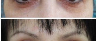
In order to prove the presence of asymmetry in the face, an image of a person’s face is taken, composed of its two right or left halves. The result is two absolutely symmetrical portraits, which often differ from the person’s real face.
We will describe the methods that are used to correct the main parts of the face.
1. Eyebrow asymmetry. The defect develops when the facial nerve, its frontal branch, is damaged. Also, its manifestation is facilitated by hyperfunction of the levator brow muscle. In order for symmetry to be restored, drugs are injected into the muscles (frontal, corrugator brow): Lantox, Botox, Dysport.2. There are numerous muscles around the mouth that work in different directions. Asymmetry around the mouth is corrected by introducing Botox, Lantox into the area of the muscle that depresses the lower lip and the area of the mouth
3. For asymmetry of the eye slits in the presence of the “big eye versus small eye” symptom, medications Lantox, Botox, and Dysport are used. A small dose of botulinum toxin can also be used; the drug is injected into the lower part of the orbicularis oculi muscle, 1 mm away from the edge of the eyelashes.
We all understand that the body of any person cannot be absolutely symmetrical, so there is no need to worry about this. But when eye asymmetry occurs, this already indicates the presence of pathology and you should definitely consult a specialist for advice.
When eye asymmetry occurs, the reasons may be the following: 1. The presence of infectious eye diseases, in this case with slight swelling of the eyes becoming larger (for example, barley or adenoviral conjunctivitis). The tumor occurs due to an attack by pathogenic bacteria, which leads to inflammation of the mucous membrane of the eye.
We invite you to familiarize yourself with Viral conjunctivitis of the eyes: causes and types, symptoms and treatment
Therapy and diagnosis are carried out only by an ophthalmologist, because self-medication can lead to worsening of the condition. 2. Injuries. It should be borne in mind that even a small injury or bruise in the eye area can cause swelling. Treatment of injuries is carried out depending on their formation, but only by a specialist. 3.
If swelling of the eye and a change in its size occurs without a reason, this situation is considered the most dangerous. This defect can lead to neurological disease or even more serious illness. 4. Bulbar syndrome is a dangerous pathology that is associated with the condition of the brain.
At the initial stage of development of the disease, asymmetry of the eyes is noted. It is at this moment that you should consult a doctor to prevent the development of a disease when one of the eyes does not function properly (for example, paralysis). Most often, in parallel with a change in the size of the eye, deformation of the eyelid, incomplete closure of the eyes, and a change in the shape of the eye occur.
Let's draw conclusions: if you have a visually visible difference in the size of your eyes, you should pay attention to the accompanying symptoms (swelling of the eyelid, discharge with purulent contents, redness of the eye mucosa). If the deformity is accompanied by attacks of pain, most often a diagnosis of “neuralgia” can be made.
All pathologies must be diagnosed by a specialist, so do not delay a visit to an ophthalmologist.
Symptoms of pathology
As a rule, with facial asymmetry, the problem is externally manifested in the fact that the right side is wider than the left. If a citizen has natural asymmetry, then the discrepancy in size between the sides can be completely insignificant in the form of 2-3 mm. This appears as a slight deformation. The patient's disturbances are expressed as follows:
- the damaged half of the face is characterized by weakness of the facial muscles and takes on the appearance of a mask;
- folds in the forehead, nose and lips are less noticeable;
- the palpebral fissure widens significantly;
- at the mouth the corners change direction (directed downwards);
- the damaged area gives the face a distressed appearance;
- the facial muscles lose their mobility, and the patient can no longer close his eyes normally or wrinkle his forehead;
- a sick person speaks poorly and cannot eat normally because it simply falls out of the mouth;
- Painful sensations often appear in the damaged area.
In children, the disorder appears when the child lies on only one side for a long time. As a rule, the face takes on a flat shape in the cheek area, and the head tilts to the injured side.
What do experts think?
Bytdaev Zaur Makhrovich, plastic surgeon, candidate of medical sciences:
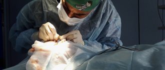
In my practice, after the recovery period there was no such problem, but immediately after the operation it can appear due to varying degrees of swelling. In order to get rid of this condition as quickly as possible, there is a list of measures that can effectively resolve the issue, including physical therapy. True, it is necessary to take into account that there is a congenital asymmetry, which is actually visible, but it can also be corrected.
Eye size difference
During the first month after surgery, differences in eye size can be considered normal. In order for everything to go smoothly, you need to strictly follow the surgeon’s recommendations and go for repeated examinations, since the doctor must monitor the rehabilitation process. If, upon completion of rehabilitation, no positive dynamics are observed, then this is considered a complication.
The first thing a patient should do in such a situation is to go to his surgeon to identify the causes and find a solution. Repeat blepharoplasty, canthoplasty, and other types of surgery may be necessary.
Aleksanyan Tigran Albertovich, plastic surgeon, candidate of medical sciences:
Eyes of different sizes occur when the operation is performed incorrectly or when the face is very uneven. I believe that first you need to choose the right surgeon, who has been performing such operations for a long time and knows how to do it, so as not to encounter unpleasant consequences in the end. Otherwise, you will have to do a second operation, which makes correcting the consequences much more difficult.
Levin Sergey Lvovich, plastic surgeon, Doctor of Medical Sciences:
Usually, in any person, the eyeball is more recessed on one side, and more protruding on the other and visually appears larger - this is a normal phenomenon. After upper eyelid surgery, the difference sometimes becomes more noticeable.
Another main factor is that after the operation the patient examines himself longer and more closely in front of the mirror and finds that asymmetry that he did not notice before. A less pleasant option is that if too much skin around the eyes has been excised, then the difference in the depth of their seating may be increased. And it can't be fixed.
Egorova Maria Vladimirovna, chief plastic surgeon at Frau Klinik:
Initially, the human skull as a whole and the eyes are asymmetrical. But almost no patient notices this. After surgery, a difference may occur due to close attention to the area as well as swelling. In general, recovery lasts 6 months; during this time, various complaints may arise regarding the operated area, which does not require additional intervention.
Different eyes are a complication if asymmetry was obtained due to incorrectly performed blepharoplasty. In some situations, correction is possible, but its feasibility can only be determined during a face-to-face consultation with a surgeon.
YouTube responded with an error: The request cannot be completed because you have exceeded your quota.
Rate this article:
- Related Posts
- How to remove seals on the lower eyelid after blepharoplasty?
- Why do my eyes itch after blepharoplasty?
- Can hollows form under the eyes after blepharoplasty?
- When is blepharoplasty considered unsuccessful?
- Why do black circles appear after lower blepharoplasty?
- Why did bumps form after blepharoplasty?
Why is one eye larger than the other?
Facial asymmetry is sometimes determined by one eye. The larger one eye is to the other, the higher the likelihood of the patient having pathology. Asymmetry especially often appears during periods of inflammation or during the development of various infectious diseases. Let's look at a few ways in which one eye can become larger than the other.
| Cause | Manifestations of the disease |
| Infection | A disease of this nature has a negative effect on the condition of the eyelid, causing swelling of the eyelids and other pathologies. A group of such diseases includes conjunctivitis or barley. Inflammation negatively affects the mucous membrane, after which one eye becomes smaller, however, this occurs almost immediately after the infection disappears from the body. It is important to support the body during this period by taking various medications. In case of complications, the asymmetry may increase. |
| Injury | A bruise or abrasion can lead to swelling of the eye, and therefore a change in the angle of vision. In this case, it is necessary to urgently visit a specialist, where a specific treatment option for the disease will be offered. |
| Bulbar syndrome | The disease develops due to complications of brain diseases. This causes deformation of the eyes, and can also cause pain and disruption of the functioning of the eye muscles. |
| A brain tumor | Malignant tumors can also act as a cause of asymmetry. If there is no reason for the problem to arise, then you should consult a neurologist. |
| Inflammation of the trigeminal nerve | Inflammation, which leads to discomfort in the ear area and severe migraines. |
Causes of eyelid asymmetry
The situation when during the first weeks and even months after blepharoplasty one eye is wider and the other is narrower is a fairly common occurrence. Behind the external manifestation of the problem there may be a whole list of reversible and irreversible changes in tissues. The main and most common cause of asymmetry of the palpebral fissures is postoperative swelling. When performing eyelid surgery, they are quite stable and can resolve within six months, sometimes longer.
Uneven accumulation of fluid in the area of the right and left eyes almost always leads to the fact that either the eyelids will be at different heights, or their shape will be deformed, or the incision on one side will be narrower than on the other. But as the swelling subsides, the eyes will also return to normal, and the problem will resolve itself. More serious causes that may require additional corrective procedures and sometimes reoperation:
- formation of persistent ptosis of the upper eyelids. Due to severe swelling, ptosis is present to any degree in the first months of recovery, but normally it should gradually go away. Alas, sometimes this does not happen completely, and if ptosis still persists within 6 months after the operation, it will not go away on its own, it needs to be removed;
- uneven facial expressions, when the muscles on one side are more developed than on the other - this feature could have been present for a long time and became obvious after surgery or developed already during the rehabilitation period;
- uneven structure of the facial part of the skull, and this applies to soft tissues and bone structures. In this situation, a small difference in the size or shape of the eyes was likely visible before surgery, but became much more noticeable after surgery;
- creating rough scars that strongly tighten the tissues of the eyelids or change the shape of the palpebral fissure. It is noteworthy that the small, barely visible difference between the heights of the upper eyelid folds may be related to which hand the surgeon has the dominant hand. Corrections make a slightly less neat cut on the left side, and vice versa - but if the scars are healed without complications, outwardly everything will look perfectly smooth;
- errors by the surgeon at the time of preparation and performance of the operation: removal of too much or insufficient amount of skin, injury to muscle fibers or tendons, incorrect suturing.
We recommend: Why does swelling under the eyebrow after blepharoplasty not go away?
How is the disease diagnosed?
To begin treating the pathology, it is necessary to conduct a diagnosis and make an accurate diagnosis. As a rule, to identify an illness, the doctor first visually examines the patient’s face for the presence of pathologies of muscles, teeth or nerve endings. After a visual examination, the specialist talks with the patient regarding hereditary pathologies, as well as the presence of serious injuries.
At the next stage, it is necessary, using a measuring device, to determine the proportions of the face and identify damage or deformation. The pathological disorder starts from 3 mm or 5 degrees. To definitively check for the presence of asymmetry, a drawing of the patient's face is performed using two right or left sides. The result is two identical images, which may have striking differences from the patient's face.
How to recognize pathology?
The main goal of diagnosing ptosis of the upper eyelid is to establish the cause that led to the development of this disease.
- Assess the position and mobility of the eyelid.
- Assess the symmetry of eye movements.
- Assess eyebrow mobility.
- Determine the size of the eyelid fold.
- Determine the strength of the muscle that lifts the upper eyelid.
- Determine the presence of strabismus, amblyopia.
- Check your vision.
- Measure intraocular pressure.
If ptosis is caused by mechanical damage, the doctor should check the bone structures for damage. To do this, you need to conduct a survey x-ray. If there is a suspicion that ptosis has appeared due to problems with the nervous system, then a computer or magnetic resonance imaging of the brain is performed, and a referral to a neurologist and neurosurgeon is given.
- General clinical blood test. It will show leukocytosis and accelerated erythrocyte sedimentation characteristic of inflammation.
- Biochemistry of blood. This laboratory test will help identify cytolytic enzymes. Their level increases if there is destruction of cells and tissues in the body.
- General ophthalmological examination. As part of it, visual acuity is measured using Sivtsev tables.
- Perimetry. This is a method for determining peripheral vision and the degree of its impairment.
- Ophthalmoscopy. Using a slit lamp, the technique examines the condition of the fundus of the eye, the exit point of the optic nerve, the vascular network, and the anterior and posterior chambers of the eye.
- Magnetic resonance and computed tomography of the head. These instrumental methods help to identify hemorrhages, foci of destruction or neoplasms in the cranial cavity.
- Measurement of cerebrospinal fluid pressure. At the same time, cerebrospinal fluid should be collected for microbiological examination.
We suggest you familiarize yourself with Drops to prevent your eyes from watering
Procedure for treating eye asymmetry
To cope with the disease and return the patient to a healthy appearance, patients use various techniques. If the pathology is not very pronounced, then there is no need to correct such a violation. Treatment is prescribed strictly on an individual basis and depends on the cause of the pathology:
- If muscle tone is affected, a course of myostimulation and a gymnastic complex for the facial muscles can help. In addition, it is necessary to carry out a massage with an emphasis on the damaged areas;
- a certain external style (hairstyle, makeup) is selected for patients, and a mustache or beard is selected for patients;
- the doctor prescribes orthodontic treatment;
- use methods of maxillofacial surgery;
- use various methods of correction using plastic surgery;
In particularly difficult cases, when the disease is severe, special neurological treatment is prescribed in a hospital with full supervision by an experienced specialist.
Eye asymmetry is a complex disease that definitely requires proper treatment. After completing the treatment procedure, it is important to adhere to the doctor’s recommendations in order to eliminate problems with possible complications and the development of other diseases associated with eye asymmetry.
To learn more about eye diseases and their treatment, use the convenient site search or ask a specialist a question.