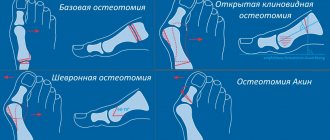
PREFACE TO LASER GRINDING OF THE FACE
I came to the clinic with problems typical for my age - thinning of the skin, decreased elasticity and turgor, fine wrinkles, dull, unhealthy complexion, which I periodically “corrected” by visiting the solarium.
Frequent sunbathing naturally worsened the condition of the skin: the skin became even thinner, dryer, flabby, with reduced tone and a large number of fine wrinkles. In general, a vicious circle. Of course, when I was offered deep laser peeling (laser resurfacing), I was afraid. Is it conceivable to practically burn the skin on your face? On the forums I read about some kind of scabs, scars and other passions. But they explained everything to me in great detail, told me about the terms of rehabilitation and the expected effect. And so, I made up my mind and took a 2-week vacation from work.
Photo before surgery
It can be seen that the skin is very uneven and uneven in color.
I had to constantly powder and brown.
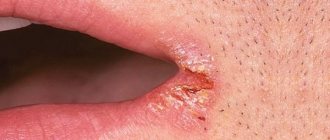
Causes of a red spot
Various marks on the surface of the skin remain after various methods of mole removal. There are several reasons for the appearance of hypertrophic scars:
- Depth and size of the removed mole. The likelihood of a bump appearing on the skin after excision of a birthmark increases in the presence of large moles and nevi.
- method . After laser coagulation, a red spot appears in rare cases, since during the procedure only the upper layer of the epidermis is affected. But after surgical removal with a scalpel, pink and red spots may remain that do not go away for 6-8 months.
- wound care rules The rehabilitation period after surgical removal of a mole is of great importance. Failure to follow the rules may cause red spots to appear. Most often they occur when the wound is exposed to sunlight.
On this topic
- Moles
All about dangerous moles
- Inna Viktorovna Zhikhoreva
- September 27, 2020
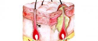
Scar stretching and inflammatory processes can also have an impact and significantly increase the risk of a hypertrophic spot.
At the same time, fibroblasts are activated, cells begin to form more actively and collagen synthesis increases. The appearance of hypertrophic scars occurs against the background of increased collagen breakdown and the development of fibrous tissue.
FIRST DAY AFTER LASER GRINDING OF THE FACE
As soon as I recovered from the anesthesia, I immediately began to look at myself in the mirror and take pictures with my phone. Under a thick layer of ointment there was a red, red face! The next day I felt a burning sensation and pain. They immediately gave me an anesthetic injection, in the evening I began to ache a little again and the injection was repeated. And so, everything was wonderful.
Photo after surgery
ABOUT THE PROCEDURE
Under the influence of the laser, the walls of the vessel “glue” together, without damaging the surrounding healthy tissue. The blood flow in the vessel stops and it is replaced by connective tissue.
Small vessels are removed almost without a trace, and large ones turn pale, significantly decreasing in size
After the procedure, slight redness of the skin remains, which disappears after about 30-60 minutes. The treated small vessel first darkens, and after about 2-3 weeks it completely disappears.
Make an appointment
FIRST WEEK AFTER LASER GRINDING OF THE FACE
They gave me painkillers, some other tablets, washed my face every 2 hours and applied magic ointments again, after which the discomfort and feeling of tightness immediately disappeared. This went on for three days. The only thing that annoyed me was the inability to wash myself! Only on the third day I was allowed to take a warm shower, avoiding my face and head.
On the fifth day after laser facial resurfacing, I was discharged. I looked like this:
At this point, brown crusts have appeared. I looked, of course, shocking, red face with brown spots. At home, I continued to wash and smear myself. Even a little more often than necessary, because cat hair was sticking to me.
General principles of treatment for keratomas
Tumors of this type can only be treated with radical methods, but this is not always necessary. Seborrheic keratoma, the follicular form, degenerates into skin cancer extremely rarely. Their development does not cause physical discomfort, so there is no need to treat them.
For patients who have such formations, doctors recommend visiting a dermatologist once or twice a year and together with him monitor the dynamics of the pathology. When the first signs of malignancy appear, it is better to remove the keratoma immediately.
Doctors can use radical treatment at the request of the patient. It occurs when the formation of a tumor creates a visible cosmetic effect that causes psychological discomfort.
THIRD WEEK AFTER LASER GRINDING OF THE FACE
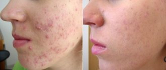
After 2 weeks, acne appeared. I was warned of a possible exacerbation (in my youth I was tormented by acne), but I hoped that this would not affect me. I started applying Metrogyl, and in three days they went away without leaving any traces.
In addition to spots and pimples, it seemed that the whole face seemed to sag and periodically swell a little.
On examination on the 10th day after laser facial resurfacing
Dermatologist-cosmetologist Elizaveta Alekseevna Gintovt:
Post-inflammatory pigmentation
Occurs after prolonged inflammation as a result of injuries, plastic surgery (if the surgeon’s recommendations are not followed), laser resurfacing, acne, or due to a reaction to cosmetics.
For some, every injury leads to pigmentation - all it takes is a cut and a dark spot will remain. This tendency often manifests itself with age, when vascular regulation is disrupted. Also, patients with dark hair and dark skin are more prone to developing congestion spots.
Melasma
Melasma can be triggered by UV radiation, pregnancy, menopause, taking hormonal and photosensitizing drugs, hereditary factors, and some gastrointestinal diseases.
Melasma happens:
- epidermal, clearer and darker: when it begins to get dark, the spots clearly appear even under a good layer of makeup;
- dermal pigmentation is lighter, but it lies in a deeper layer of the skin;
- mixed pigmentation is formed due to the accumulation of pigment cells in the dermal and epidermal layers.
Precautions against pigmentation:
1. Use sunscreen. Guaranteed protection is only a completely UV-blocking cream with zinc, factor 100. There are no guarantees when applying sunscreen with factor 50. People prone to pigmentation should use sunscreens from the beginning of March. Even in Moscow there is already a danger during this period, so in the spring we do not carry out photosensitizing procedures (laser resurfacing, etc.).
Some patients complain that in sunny times they do not take off their hat and only tan their bodies, but the pigment appears. Yes, this is possible, since melagenesis starts in the body as a whole.
2. Do not abuse the solarium. Covering your face and sunbathing only with your body will not protect you from pigmentation, since melanin production is a general process.
3. Do not sunbathe when using drugs that provoke photosensitivity. A group of tetracycline antibiotics increases photosensitivity. Hormonal drugs and oral contraceptives often act as photosensitizers. Therefore, you need to carefully study the instructions: as a rule, similar properties are indicated. If you take such drugs and then go to a sunny country or sunbathe even in the middle zone, pigmentation will appear if there is a tendency, and if pigmentation already existed, then it will intensify several times. You also need to be careful with aggressive cosmetics: peelings, retinol-based products.
Remedies to combat pigmentation at home
To combat pigmentation, retinoids, preparations based on azelaic acid (for example, Skinoren), professional creams with glycolic acids (for example, creams from SkinCeuticals, Dermaceuticals) are used. These drugs help with any type of pigmentation with proper and long-term use.
Hydroquinone is currently not approved for sale because it is very toxic, although it is effective even in the fight against dermal melasma.
Anti-pigmentation products from a cosmetologist
For post-inflammatory pigmentation
you need to choose a course of procedures so as not to get new post-inflammatory pigmentation: already because of attempts to get rid of it. Sometimes it is necessary to first carry out mesotherapy, plasma therapy, improving the quality of the skin and relieving inflammation, and only then remove the pigment.
Dealing with post-traumatic pigmentation is difficult. Therefore, if you notice that redness persists for a long time after an injury, go to a cosmetologist.
With dermal pigmentation
You may need a course of procedures that will have to be repeated next year, or maybe within 2-3 years.
Different lasers may be needed to combat different age spots. Only the doctor can make the choice. You can use V-Beam when the pigment spot is also based on a vascular component. Sometimes we use a CO2 laser, sometimes a thulium laser. I like the Fraxel thulium laser - it is non-aggressive and effective.
Epidermal spots go away quite quickly, sometimes one Fraxel procedure (using a thulium laser) is enough. Dermal melasma is more difficult to deal with: we can make the spot paler, but in spring and summer the pigment may return and the course of procedures will have to be repeated.
Phototherapy can also be used, but, unfortunately, the effectiveness of this technique is much lower than that of laser.
Peels based on retinol and glycolic acids are also used. At the same time, biorevitalization will be useful for strengthening and moisturizing the skin.
Also read:
“Painting bags on cheekbones: how to get rid of them?”
“Cosmetology in the summer: what is possible”
“The result of aggressive resurfacing... without heavy rehabilitation”
“Micolblading of eyebrows: from the creators of the technique”
"Complications after Botox"
This is not a public offer! There are contraindications. Before use, consultation with a specialist is required.
Care instructions
Strict adherence to certain rules will help speed up the pace of recovery, as well as avoid complications and relapses.
After removal of papillomas you cannot:
- Injure the wound surface (avoid any mechanical damage, wearing tight clothes or underwear);
- Cover the wound with an adhesive plaster (free access of air helps speed up the process of regeneration and scab formation). It is allowed to apply a bactericidal patch when going outside;
- Stay in the open sun for more than 10 minutes (the same bactericidal patch will help protect the wound from the aggressive effects of ultraviolet radiation). If the tumor is located on the eyelid, wear sunglasses;
- Keep it wet for 24 hours, and even more so wash the wound with soap or other hygiene products (you need to wash very carefully, also do not rub the wound surface with a towel, just blot excess moisture with it, this will be enough);
- Visit solariums, go to baths, swim in pools and open-air reservoirs for at least 3 weeks. Also, during this time it is not recommended to use decorative cosmetics.
- Tear off the crust from the surface of the wound (this will prolong the recovery process).
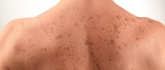
Types of pigment spots
Skin tone depends on melanocytes - special cells. When their work is disrupted, the epidermis becomes covered with pigment spots of several forms. Here are their varieties:
- lentigo - these round spots have a clear outline and reach up to 50 mm. Depending on the age at which they appear, they distinguish between juvenile and senile lentigo;
- chloasma is an area where there is more melanin than in other places. The shade and borders are uneven. The size may be quite large;
- nevus, colloquially a mole, is a flat or protruding pigmented formation that has different shapes, sizes and shades;
- ephelides, simply freckles - many small round yellow-brown spots;
- Brocca's meloderma - brown spots on the lips, sometimes on the nose. Usually appear as a consequence of stress.
Meloderma Brocca
Keratoma removal methods
Removal of keratomas can be done in different ways:
- laser;
- liquid nitrogen;
- electrocoagulation;
- radio wave method;
- traditionally with a scalpel.
The choice of method is made strictly on an individual basis. The patient’s age, size and shape of the keratoma, and available technical capabilities are taken into account.
If there are signs of malignancy, the patient will be offered laser or traditional surgical treatment. Only in this case is it possible to obtain biological material suitable for histology. This is the most informative analysis that allows you to determine the stage of development of a malignant tumor and find out how far the process of metastasis has gone.
Removal with a scalpel
Traditional surgery is performed under local anesthesia. The surgeon uses a scalpel to cut out the keratoma itself and the tissue adjacent to it. Then the wound is sutured, the seams are treated with an antiseptic solution, and a sterile bandage is applied over them. The recovery process lasts two weeks. In the first seven days, the wound is treated and bandaged three times a day. The patient is undergoing a course of antibiotics. They help prevent bacterial infection of the surgical site.
Why red dots appear on the body: what is it or is it all about angioma
Many people are particularly interested in red dots on the body. Is this a sign of a serious illness or a completely harmless formation? Red moles are benign tumors that develop from lymphatic or blood vessels.
Angioma is the correct name for this phenomenon. One of the most mysterious tumors has long haunted the minds of scientists studying this strange formation. Are red dots on the body dangerous? Should they be removed? Let's figure it out.
- What is angioma
- Possible reasons for the appearance
- How to get rid of red moles
- Modern removal methods
- Home Remedies
- Red dots on a child's body
- Preventive recommendations
What is angioma
A benign tumor is formed by the expansion of one or more capillaries. By their nature, these are nothing more than modified vessels.
This statement is easy to verify. Apply pressure to the angioma. The formation will quickly turn pale, and after a few seconds it will return to its previous appearance. There is no mystery here - the blood has filled its place again. This process is clearly visible on large tumors.
Crimson dots have different sizes - from 0.5 mm to several centimeters. The more dilated the vessel is, the larger the angioma.
Sometimes there are several affected areas along the blood vessel. The disease is called hemangiomatosis. Large or small dark red spots are visible along the entire leg or arm.
Types of red dots:
- capillary hemangioma. The simple form is characterized by the usual expansion of capillaries, small red dots on the body look like moles. Location: face, torso. Color – blue-crimson, dark red. The formations look like small, individual points on the body;
- branched hemangioma. A benign tumor resembles a swelling consisting of pulsating, highly dilated vessels. After pressing on the area of the hemangioma, the blood immediately leaves and then also quickly fills the vessel. Color – deep red;
- cavernous hemangioma. The dark blue formation consists of large cavities filled with blood. This type is often found on the face and worsens the appearance. The formation is covered with thin skin. The nature of cavernous hemangioma explains why such cavities are detected on the spleen, head, uterus, and liver.
What do red dots on the human body mean and is angioma dangerous? Doctors reassure that a benign formation can exist for years without causing harm. There have been virtually no cases of angiomas degenerating into malignant tumors.
Read about methods for removing a spine on your foot on our website.
Read all about erythema annulare in children and adults in this article.
Possible reasons for the appearance
It is not for nothing that dermatologists from different countries consider angioma to be a completely unstudied phenomenon. The exact reason that causes the appearance of deep crimson, crimson or dark blue tubercles on various parts of the body is still not known.
Why do red dots appear on the body of an adult? There are several versions. Scientists' guesses are based on:
- studying the clinical picture;
- time of occurrence of vascular formations;
- depending on the size, their appearance, and the condition of various organs and systems.
The main reasons for the appearance of small red dots on the body:
- diseases of the liver, pancreas. Individual elements of burgundy, crimson, and reddish hue are always found in the upper part of the body. Deterioration of the pancreas and liver causes the appearance of new red moles. When the functioning of these organs is normalized, angiomas completely (partially) disappear or decrease;
- diseases of the digestive tract. One of the probable reasons. Red moles are located in the same way as in liver pathologies. The size, number, and color of benign tumors vary depending on the condition of the gastrointestinal tract;
- microtrauma of the skin. The version has a right to exist;
- lack of vitamin K and ascorbic acid. A deficiency of these important components leads to damage to the vascular walls. The result is dilation of blood vessels in a certain area, the formation of a benign tumor;
- allergic reactions. There is also such an assumption. Angiomas occur as a reaction of the body to an irritant - drugs, dust, household chemicals, pollen, food, cosmetics and other allergens.
Note! Most researchers believe that versions of pathologies of the liver and digestive system are the most plausible.
How to get rid of red moles
Is it necessary to do this? Benign formations usually do not bother the owner and do not require drug treatment. Small separately located elements should not be removed.
Perhaps, having improved the functioning of the digestive tract, treated the liver and pancreas, and cleansed the body of toxins, you will find that the hemangiomas have become smaller or disappeared. Sometimes purple formations disappear as suddenly as they appeared.
Consult a dermatologist:
- the doctor will prescribe an examination of various organs and systems;
- after a conversation with the patient, he will analyze at what period the red moles appeared, whether there were any malfunctions in the body at that time or the patient was sick with something;
- Based on the test results and clinical picture, the specialist will decide whether the red dots need to be removed or not.
It is necessary to get rid of angiomas in several cases:
- a cosmetic defect disfigures the face or another open area of the body;
- a benign tumor is located in an inconvenient place and is constantly subject to friction;
- There is a danger that you can accidentally tear off and injure a red mole.
Modern removal methods
Methods for removing benign vascular formations are similar to methods for removing warts. The dermatologist will determine which method to choose.
Removing hemangiomas on the face is also not a cause for concern. Modern cosmetology has effective techniques that leave barely noticeable marks on open areas of the body.
Basic methods of cosmetic surgery:
- laser removal. An effective method: the use of laser leaves almost no traces and helps get rid of hemangiomas even on the face;
- cryodestruction. Liquid nitrogen acts on red dots and freezes them. After a while, the crust disappears, leaving an inconspicuous scar at the site of the operation. Sometimes there is some pain in the postoperative period;
- surgical excision. The method is effective for removing large formations. The disadvantage is a noticeable scar. Not recommended for removing angiomas on the face;
- electrocoagulation. The high-frequency current only affects the mole and burns it out. The crust disappears after 10 days;
- radiosurgery method. One of the most effective ways to combat various formations on the skin. The radioknife cuts out the tumor, prevents the development of bleeding, and disinfects the wound. There are no traces after the operation. The short postoperative period proceeds without complications.
How to get rid of acne on the back and chest? Learn effective methods.
The causes and treatment of keratosis of the skin are written at this address.
If you go here https://vseokozhe.com/borodavki/papillomy/kak-ybrat.html you can find out everything about the treatment of human papillomavirus.
Home Remedies
Some publications publish folk recipes for hemangioma. Dermatologists believe that only radical measures are effective.
Please pay attention to these cautionary tips. The price of the issue is health.
Remember:
- it is impossible to remove reddish, purple moles with pineapple, lemon, onion juice, garlic, red radish, and dandelion compresses;
- remember the vascular nature of hemangioma. Forcing part of a blood vessel to dissolve or trying to lighten it is, to put it mildly, unreasonable;
- the use of harsh, acidic foods can injure the lining of the vessel. You will get nothing but bleeding;
- no one can guarantee that after treatment with irritating substances, infection will not penetrate into the damaged vessel walls. The consequences can be very serious.
Red dots on a child's body
Angiomas often form in children of different ages. Poor environment, stress, low-quality products provoke diseases of the digestive system, pancreatitis, hepatitis, and other misfortunes.
The appearance of moles of a reddish, purple hue is a reason to visit a dermatologist. The doctor prescribes an examination by a hepatologist or endocrinologist. It is possible that diseases that provoke the development of hemangiomas will be discovered.
Should I touch red moles or not? Most dermatologists have a negative attitude towards removing angiomas in children.
Do nothing about hemangiomas if they:
- do not rub against clothes;
- are not at risk of injury;
- are not a noticeable cosmetic defect.
Helpful Tips:
- Remain calm when you discover strange purple spots on your baby's body. Red moles do not pose an oncological danger;
- monitor the condition of benign tumors, note during which periods they became more or less numerous. Perhaps the formations will disappear on their own. Based on the records, the most likely cause of angiomas in your child will be clear;
- whether new formations will appear on the body or not - largely depends on lifestyle, the condition of blood vessels, the presence of diseases of the gastrointestinal tract and endocrine system; take an interest in your child’s health – it will be easier for you to respond to the manifestations of any disease.
Preventive recommendations
Based on the likely causes of the formation of red blood spots, the following can be recommended:
- monitor the functioning of the digestive organs, liver, pancreas;
- get examined in a timely manner, treat acute and chronic gastrointestinal pathologies;
- eat a varied diet, get enough vitamins, including K and C. They are found in cabbage, lemons, black currants, spicy herbs, rose hips, plums, and prunes. Don't forget about red peppers, sea buckthorn, leafy green vegetables, and fresh fruits.
These tips will also be useful:
- strengthen your immune system;
- monitor the condition of the vascular system;
- eat right.
Now you know that red dots are not as dangerous as they might seem at first glance. The more questions asked, the more detailed the problem can be understood.
Below is a video from which you can learn even more details about angioma:
Popular questions
What does skin treated with laser for hyperpigmentation look like?
At first the spot becomes darker. Then it peels off for several days, becoming lighter in the process, until it disappears completely.
The skin may peel for a while
How long is the entire course?
This is determined by the characteristics of the spot, intensity, and depth of melanin. A dermatocosmetologist will assess the number of procedures during a personal consultation.
Is it possible to remove freckles this way?
Of course, like any other types of age spots. And thanks to its safety, the method is also suitable for treating the décolleté and periocular area.
What is the difference between laser therapy for hyperpigmentation and chemical peeling?
This peeling does not distinguish between cells oversaturated with melanin and the rest of the skin. It just deletes it all. And this is very harmful.
What is the rehabilitation period?
He's gone. Just don't wear makeup on the day of the procedure.
What time to choose for laser depigmentation?
You can choose any season for this. The main thing is to use a very good sun protection cream.
This procedure can be carried out at any time of the year.
When should the procedure not be performed?
Contraindications include some diseases, elevated body temperature, pregnancy, and lactation.
What is the minimum age for the procedure?
This procedure can usually be performed on people over 10 years of age unless otherwise directed by a doctor.
The procedure can be performed even on children
After mole removal
The best way to remove a nevus today is laser. This method is bloodless, practically painless, after the evaporation of skin formations there are no scars left, and nearby tissues are not subject to traumatic effects.
After destruction of nevi, in most cases no complications arise. If, after removing a mole, no matter how, a red spot remains, this indicates that the healing process (granulation) is in full swing. After some time, this redness goes away on its own, and no traces of the operation remain (sometimes a barely noticeable spot can be observed).