Ptosis is a common pathology, which is manifested by a drooping of the upper eyelid towards the edge of the iris by more than 2 mm. The disease is expressed not only visually, but can also cause deterioration in visual ability. Cosmetic surgery - blepharoplasty - will help eliminate the defect.
In this article
- Causes and symptoms of ptosis
- Types of Upper Eyelid Ptosis
- Congenital blepharoptosis
- Acquired ptosis
- Blepharoplasty: indications and contraindications
- Types of blepharoplasty: how to choose?
- Preparing for surgery
- Blepharoplasty: stages
- Rehabilitation
- Complications
- Cost of blepharoplasty
Causes and symptoms of ptosis
Upper eyelid ptosis or blepharoptosis can be expressed by the following symptoms:
- noticeably drooping upper eyelid;
- with bilateral lesions, sleepy facial expression;
- rapid fatigue of the visual organs during exercise (reading, working on a computer, watching TV, etc.);
- excessive lacrimation, discomfort, pain;
- discomfort when closing the eyelids;
The mechanics of development of the above-described symptoms directly depends on the tone, contractions and width of the palpebral fissure during the motor function of the upper eyelid.
Thus, the levator palpebral muscle controls its vertical position. The orbicularis muscle allows you to close the eye. And the frontalis muscle provides compression and contraction of the eyelid when looking up.
Tone and contraction occur under the influence of nerve impulses that arrive to the frontal and circular muscles from the facial nerve, equipped with a nucleus located in the stem section on the corresponding side of the brain.
The muscle that raises the upper eyelid is innervated by a group of neurons - the left and right bundles of the caudal nucleus of the oculomotor nerve, which is also located in the brain.
Symptoms of ptosis
The main clinical manifestation of the disease is drooping of the upper eyelid , which leads to partial or complete closure of the palpebral fissure. At the same time, people try to tense the frontalis muscle as much as possible so that the eyebrows rise and the eyelid stretches upward.
For this purpose, some patients throw back their heads and take a specific pose, which in the literature is called the stargazer pose.
A drooping eyelid prevents blinking movements, which leads to soreness and eye fatigue. Reduced blinking frequency causes damage to the tear film and the development of dry eye syndrome. Infection of the eye and development of an inflammatory disease can also occur.
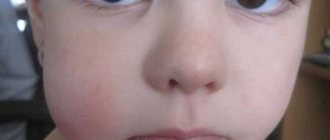
Features of the disease in children
Ptosis is difficult to diagnose in infancy. This is largely due to the fact that most of the time the child sleeps and has his eyes closed. You need to carefully monitor the baby's facial expression. Sometimes the disease may manifest as frequent blinking of the affected eye during feeding.
At an older age, ptosis in children can be suspected by the following signs:
- While reading or writing, the child tries to throw back his head. This is due to the limitation of visual fields when the upper eyelid droops.
- Uncontrolled muscle contraction on the affected side. Sometimes this is mistaken for a nervous tic.
- Complaints about rapid fatigue after visual work.
Cases of congenital ptosis may be accompanied by epicanthus (a fold of skin hanging over the eyelid), strabismus, damage to the cornea and paralysis of the extraocular muscles. If ptosis in a child is not corrected, it will lead to the development of amblyopia and decreased vision.
Types of Upper Eyelid Ptosis
Ophthalmologists divide ptosis according to the type of localization into unilateral (occurs in 70% of cases) and bilateral. Also, such a disease of the upper eyelid can be true and false. The second type may be associated with the presence of excess eyelid skin and subcutaneous tissue, endocrine pathology of the visual organs, eyelid hernia, loss of elasticity of the eyeballs, and strabismus.
In addition, a distinction is made between pathological and physiological drooping of the upper eyelids. The first occurs in connection with mechanical injuries, inflammatory processes of motor muscles, eyeballs or brain membranes, as well as anomalies at various levels (hemispheric, supranuclear, nuclear) in the nervous system after heart attacks and brain tumors, damage to the brachial plexus and others.
Physiological drooping of the upper eyelids can be associated with the sympathetic system, retina, hypothalamus, occipital and temporal cortex. Then the muscle tone and width of the palpebral fissure will depend on the emotional state of the person - fatigue, reaction to pain, surprise, anger and others. In this case, eyelid ptosis is short-lived.
According to the degree of the pathological condition in practice, ptosis of the upper eyelid is:
- partial - the upper eyelid covers only 1/3 of the pupil;
- incomplete - the eyelid covers 2/3 of the pupil;
- full - the eyelid completely covers the pupil.
Due to development, pathology is divided into congenital and acquired.
How to identify blepharoptosis
History taking
- Drooping eyelids (symmetrical bilateral minimally expressed drooping of the eyelids with full excursion of the upper eyelids. Functionally, the upper eyelid is complete, but aesthetically does not satisfy the patient).
- Patients appear sleepy or tired.
- Patients may complain of blurred vision, increased lacrimation, and a decrease in the upper field of vision.
- Difficulty with activities of daily living such as driving, reading, and climbing stairs.
- Complaints of headache caused by tension in the forehead due to excessive use of the forehead muscles when trying to indirectly lift the eyelids and raise the eyebrows.
- The patient can lift the eyelid with a finger or the entire eyebrow to improve vision.
Clinical measurements
Normally, the upper eyelid usually covers 1 mm of the cornea, and the lower eyelid usually lies at the junction of the cornea and sclera3.
The following measurements are recommended to diagnose ptosis13:
- Palpebral fissure height: Defined as the widest distance between the edges of the upper and lower eyelids when viewed straight on, and usually ranges from 8–11 mm.
- Marginal reflex distance: most accurate for measuring ptosis. The patient looks into the distance while looking straight ahead. The distance is usually 4–5 mm and does not depend on the position of the lower eyelid.
- Upper eyelid crease: The distance from the upper eyelid crease to the edge of the eyelids is about 8 mm in men and 9–10 mm in women. The upper fold of the eyelid is absent in congenital ptosis and increases in aponeurotic ptosis.
- Levator Function Test: This test evaluates the pulling strength of the levator muscle. The movement of the upper eyelid in downward and upward gaze is measured, while the brow/frontalis muscle is fixed with the hand to avoid its natural movement. Normal function is greater than 11 mm, greater than 11 mm is considered “very good”, 8–10 mm is “good”, 5–7 mm and less than 4 mm is considered “poor”.
Based on these measurements and clinical examination, ptosis can be classified as mild (1–2 mm), moderate (3–4 mm), and severe (>4 mm)
Visual inspection
- Palpate the eyelids and orbital margin.
- Assess any clinical signs of relative proptosis or enophthalmos in each eye.
- Inspect eyebrows at baseline.
- Note the position of the patient's head; chin position. Patients may tilt their head back.
- Examination of the pupil. Differences in pupil size and iris color may rule out Horner syndrome.
- Presence of strabismus.
If symptoms are present, the cosmetologist should refer patients to ophthalmologists.
Congenital blepharoptosis
The congenital form of the disease is less common in ophthalmological practice than the acquired form. Its reasons may be:
- Marcus syndrome is a drooping eyelid that is relieved by opening the mouth, chewing, yawning, or shifting the lower jaw to the opposite side. This is a syndrome with disruption of the nucleus of the oculomotor and trigeminal nerves.
- Congenital myasthenia or pathology of the levator innervation.
- Horner's syndrome is ptosis with accompanying constriction of the pupil, a more significant deepening of the eyeball, and dilation of the conjunctival vessels.
- Neural etiology with congenital paresis of the third pair of cranial nerves.
- Duane's syndrome is a rare congenital form of strabismus with a lack of motor ability of the eye to move outward.
- Isolated ptosis of the upper eyelid is caused by abnormal development or complete absence of development of the levator or its tendon. This congenital form of the disease is most often hereditary and, as a rule, is bilateral.
Treatment of blepharoptosis with folk remedies
In order to fully get rid of the aesthetic and physiological shortcomings of ptosis, traditional medicine will not be enough. Methods can only be determined by the treating ophthalmologist and prescribed as supportive therapeutic manipulations.
Among the most commonly used types of folk remedies in the treatment of blepharoptosis are:
- The use of medicinal decoctions as compresses and ice - their base requires chamomile, sage, birch or parsley. Cotton pads with a decoction are placed on the eyes, or the eyelids are wiped with an ice cube once a day after washing.
- Applying tonic or anti-inflammatory masks to the skin - masks are prepared using medicinal herbs and their oils (thyme, rosemary, lavender) and applied to the entire face and eyes for 20-30 minutes. After this, the remnants of the mask are washed off.
Through such procedures, the condition of the eyelids improves in many ways: their tone increases and the risk of additional eye pathologies decreases. In general, it is impossible to cure ptosis of the upper eyelids at home and with folk remedies. All of the listed home therapy options only weaken the symptoms of the disease, stabilize the condition, but are not able to eliminate the cause.
Acquired ptosis
Acquired ptosis or drooping of the upper eyelid is often unilateral. The disease occurs due to levator paresis or palsy. This may be a consequence of mechanical damage, tumors, age-related changes in the body, or diseases such as herpes zoster, stroke and others.
The most common forms of acquired eyelid drooping include:
- Myogenic - manifests itself in muscular dystrophy, myasthenia gravis, blepharophimosis, or is a consequence of ocular myopathy.
- Aponeurotic - most often associated with dystrophic age-related changes and weakness of the muscle aponeurosis. Less often it becomes a consequence of mechanical damage and trauma, and is treated with corticosteroid-type drugs.
- Neurogenic - the result of abnormal innervation of the oculomotor nerve in Horner's syndrome, aplasia syndrome, paresis, ophthalmic migraine, diabetic neuropathy or stroke.
- Mechanical - can be caused by severe swelling, inflammatory processes, isolated levator pathology, abnormal changes in the orbit (tumor, damage to the anterior orbital region, etc.), unilateral facial dystrophy after a stroke.
Why does prolapse occur?
Drooping eyelids can occur due to many reasons. The causes of drooping eyelid, which is congenital, are often associated with a hereditary factor. If one of the parents had drooping eyelids, then there is a possibility that the child will also have this disease. As a rule, the causes of congenital drooping of the upper eyelid are associated with underdevelopment of the muscle that ensures the elevation of the upper eyelid. Also, the congenital type of the disease may be associated with pathology of the nucleus of the oculomotor nerve , with weakness of the superior rectus muscle of the eye. In more rare cases, congenital drooping of the upper eyelid is associated with some other pathologies ( alpebromandibular syndrome , abnormally short palpebral fissure ). Treatment of drooping eyelid in this case is carried out only after an accurate determination of the cause that provoked this disease.
Acquired drooping of the lower and upper eyelids is more common. In this case, depending on the cause of the disease, ptosis is divided into several types. The reason must be taken into account during diagnosis, since the treatment of lower eyelid drooping depends on this. The fact that the patient requires correction of the upper eyelid is sometimes noticeable even from the photo.
Neurogenic ptosis is a consequence of paralysis of the oculomotor nerve. This symptom is observed in patients with tumors , diabetic neuropathy , and intracranial aneurysms .
The myogenic form of this symptom develops in patients with myasthenia gravis . In this case, bilateral prolapse may occur. Due to the strain on the eyes, the patient develops double vision.
Aponeurotic ptosis is typical for older people, in whom the levator palpebral muscle gradually moves away from the plate to which it is attached. As a result, the tension of the eyelid and its fixation disappear. This symptom can develop in a person who has suffered trauma.
The mechanical form of the symptom is a consequence of scarring or swelling, as a result of which the eyelid is horizontally shortened.
False ptosis is also determined, the cause of which is too large skin folds above the eye. This phenomenon can also occur in people with strabismus and hypotonia of the eyeball.
Blepharoplasty: indications and contraindications
It is important to understand that blepharoplasty is a surgical intervention, usually of a cosmetic type, but it can also be of a therapeutic nature. The operation is performed to change the shape of the eyelid, helps eliminate its overhang, and relieves the patient of ptosis.
Experts identify the following indications, in addition to the overhanging upper eyelid, for blepharoplasty:
- the patient has large bags under the lower eyelids;
- severe form of blepharoptosis, in which a person experiences discomfort;
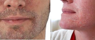
- the appearance of severe dark circles under the eyes (it is important to remember that this symptom is characteristic of many diseases, so you first need to establish the cause of its occurrence);
- wrinkles at the outer edges of the eyes.
Before the operation is performed, the doctor identifies the causes of ptosis. For example, the congenital form of the pathology looks symmetrical, and its cause is abnormal muscle development. Therefore, as a consequence, “lazy eye” syndrome appears. If blepharoptosis of the upper eyelid is acquired, it is usually the result of a muscle strain, injury or nerve disease.
Contraindications in the presence of which blepharoplasty should not be performed include:
- pronounced pigmentation of the skin in the area around the eyes;
- tattooing in the eyelash area;
- systemic diseases during the period of decompensation;
- acute forms of inflammatory diseases of any origin;
- menstrual period in women.
Treatment of the defect
Initially, therapy for drooping eyelids is carried out with the aim of eliminating the very cause that influenced it, and then the cosmetic defect is corrected.
If the drooping eyelid is of a neurogenic nature, the main cause is also treated first, and then local physiotherapeutic treatment is used as an addition: paraffin therapy, galvanization, UHF.
Congenital drooping of the eyelid, or in the case where conservative treatment has not given the expected results after 9 months, surgical ophthalmological intervention is performed. It is possible to quickly eliminate a birth defect at certain periods of life. If the eyelid partially covers the pupil, then the operation is performed between the ages of 13 and 16 years. If the eye is completely closed due to the fact that amblyopia may develop, the patient is operated on in preschool age.
The operation to eliminate this defect is aimed at reducing the length of the eye muscle, which is responsible for raising the eyelid. This procedure is used for congenital pathologies. To eliminate acquired eyelid drooping, the levator aponeurosis is shortened.
In case of congenital pathology, the levator is isolated, plication is performed by excising the muscle, or a duplication is created. In case of pronounced drooping of the muscle responsible for raising the eyelid, it is sutured to the frontal muscle.
During standard surgery to eliminate an acquired defect, a small area of the upper eyelid is excised. In addition, the aponeurosis is resected, and its lower edge is fixed to the cartilage of the eyelid.
During eyelid surgery, upper blepharoplasty is performed in parallel.
Best materials of the month
- Why you can't go on a diet on your own
- 21 tips on how to avoid buying stale food
- How to keep vegetables and fruits fresh: simple tricks
- How to curb your sweet cravings: 7 unexpected products
- Scientists say youth can be extended
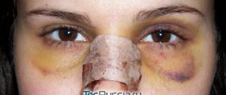
After surgery, swelling appears. To reduce it slightly, it is necessary to apply cool compresses during the first 24 hours after the intervention. This could be a dry but cold towel, or any freeze-dried product. Every day the swelling will subside, and the eyelid will return to its usual appearance and color. All this will take approximately fourteen days. After this, the person will be able to resume his usual activities and work.
However, after the operation, not only swelling may appear, the eyelid may not close completely. Until everything returns to normal, the mucous membrane of the eye must be constantly moisturized. For this purpose, special drops and ointments are used. They should be prescribed by the attending physician after the operation.
Types of blepharoplasty: how to choose?
For a patient with a problem with a drooping upper eyelid, a specialist can offer several surgical options. The choice of method for carrying out the procedure will depend on the physiological characteristics of the person, the degree of impact of the pathology on the visual organs and the nature of its origin.
The following types of blepharoplasty are distinguished:
- Traditional upper eyelid surgery - the surgeon removes a little fat layer through a micro-incision, and then sagging skin. Subsequently, the skin is fixed with anatomical “anchors”.
- Laser correction - using a laser beam, a specialist makes several miniature incisions and removes not only hernias, but also existing fat deposits. The peculiarity of this type of blepharoplasty is minimal trauma.
- Transconjunctival intervention is a sutureless technique that is indicated for patients with slight drooping of the upper eyelid and congenital type of ptosis. That is, in the case when the regenerative abilities of the patient’s body are normal.
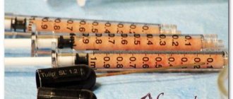
- Injection blepharoplasty - by injecting special substances that help get rid of drooping upper eyelids and dark circles under the eyes.
It is important to understand that the choice of type of operation is made individually after examining the patient.
Breast ptosis - symptoms and treatment
The method for eliminating ptosis depends on its degree.
Grade I ptosis and pseudoptosis can be removed by installing implants. The skin will straighten, volume will be added, and the breasts will take their previous shape.
A small degree of ptosis can also be solved by endoscopic lifting, which is performed using small incisions in the armpit.
Ptosis of degrees II and III must be corrected by removing excess excess skin. This operation is performed using sutures. They come in three types: areola stitch, vertical stitch and anchor stitch. Which suture the doctor chooses depends on the individual characteristics of the patient. Moreover, if the patient does not have enough volume, it can be added by installing implants. Thus, two operations are performed at once: tightening and augmentation.[5]
The periareolar method of surgery (using a suture along the areola) leaves the least traces, but is used only for a small degree of ptosis. If the breasts are very sagging or there is asymmetry, this method of lifting will not be suitable.
The vertical lift method is desirable for patients with severe ptosis, as well as for those who need to add volume with an implant. However, in some cases, for example, when it is necessary to move the nipple, a vertical seam will not work.
The anchor method of lifting allows you to solve the widest range of problems:
- greater degree of ptosis;
- asymmetry of the mammary glands;
- displacement of areolas;
- insufficient or excessive volume;
- sagging of the lower part of the breast.
However, this method has the most pronounced seams.[6][7]
Ptosis is sometimes combined with breast hypertrophy, that is, with its too large size. This can be either a consequence of breastfeeding or the girl’s hormonal background. In this case, the plastic surgeon performs reduction mammoplasty, that is, it removes not only excess skin, but also reduces the size of the mammary gland.[8]
Postoperative complications
Complications after a breast lift are divided into natural and pathological.
Natural complications:
- Swelling after surgery. On average, they go away in 3-4 days, maximum in a week. In some cases, the surgeon prescribes additional medications to the patient that relieve inflammation and swelling.
- Chest pain. They may bother the patient for the first couple of weeks after surgery, so the doctor usually prescribes pain medication.
- Decreased breast sensitivity. This complication is also common in the postoperative period and goes away after a couple of weeks.
- Scarring. The first few months they are quite clearly visible. It is at this time that it is not recommended to be in the sun, as the seams may become pigmented. However, after a year they are completely healed. If necessary, they can be corrected later using a laser.
Pathological complications:
- Minor bruising and hematomas are normal after surgery. However, large hematomas can be dangerous and require surgical removal. Most often, they occur when the patient does not comply with postoperative recommendations, in particular, for the first time you cannot lie on your side and stomach.
- Seams coming apart. Normally, the suture heals within 7-10 days, after which a thin scar remains. However, if the edges do not adhere tightly to each other, suppuration may begin between them, or they may separate. This complication appears both due to insufficiently professional work of the doctor, and in connection with the individual characteristics of the body: impaired blood circulation and weakened immunity.
- A fistula is a hole that independently forms in the skin during a purulent process. It occurs when an infection occurs in the connective tissues of the breast during surgery.
- Bad result. The reason for this may be either the lack of professionalism of the plastic surgeon or the individual characteristics of the patient, which were revealed during the operation.
If after surgery the patient regains weight and then suddenly loses it, or gives birth to another child, a repeat operation may be required.
Complications arise in exceptional cases. Modern diagnostic methods make it possible to identify all the characteristics of the body even before surgery and correctly correct all the nuances. Moreover, modern types of anesthesia and surgical technologies have minimal impact on the patient's health. The most important thing is to choose a doctor who inspires trust.
Preparing for surgery
Before starting the procedure, the specialist conducts a comprehensive examination of the patient. It is important for the doctor to know whether he has had chronic pathologies, whether there are diseases in the active stage, whether surgical interventions have been performed previously, what is the plasticity of the skin and the depth of wrinkles, and whether there are allergies to specific medications.
In addition, a patient with a drooping upper eyelid must undergo a number of instrumental and laboratory tests, including an ECG, general urine and blood tests, blood biochemistry, and a clotting test.
Most often, blepharoplasty is performed under local anesthesia, since general anesthesia is a serious stress for the body.
If the operation is complex, not only on the upper but also on the lower eyelid, the patient is offered general anesthesia. Because this type of procedure is more traumatic.
How does prolapse manifest?
Eyelid ptosis occurs in both children and adults. Ptosis of the upper eyelid can be congenital and acquired (manifests due to certain reasons). Depending on the severity of the disease, complete (the pupil is completely covered by the upper eyelid), partial ptosis of the upper eyelid (a third of the pupil is closed from above) and incomplete ptosis (two-thirds of the pupil is closed) are determined.
There is also a classification depending on the degree of damage: with a unilateral form of the disease, only ptosis of the left or right eyelid is noted. If it is bilateral , both eyes are affected at once. Sometimes ptosis of the lower eyelid is diagnosed.
This pathology occurs not only in adults: ptosis in children is a fairly common phenomenon.
If a person develops ptosis of the eye, he may think that this is not a particularly significant problem. Ptosis of the left eye or right eye initially seems to be only a cosmetic defect. But we should not forget that such a pathology is a serious obstacle to vision. This does not allow the patient to fully see, therefore, he has to constantly strain his eyebrows to free the eye from such interference. In some cases, you even have to tilt your head back to see normally. Therefore, with such a disease, a person’s quality of life noticeably deteriorates.
Patients often develop irritation and experience rapid eye fatigue, as the person is forced to constantly make an effort to gain a normal view. Often such people develop strabismus , as well as double vision . A person sometimes cannot completely close his eye, and his upper eyelid mobility is limited. The eyes often feel dry. In the damaged organ of vision, vision gradually deteriorates and amblyopia - a deviation of vision to one side.
Blepharoplasty: stages
Upper eyelid correction surgery includes several stages:
- applying markings to the eyelid using a marker;
- applying a plate to the visual organs to protect against injury;
- an incision in the eyelid in the area designated by markings;
- moving the skin of the eyelid to the side to remove the fat layer;
- removal of excess skin;
- suturing;
- treatment with an anesthetic anti-inflammatory solution and application of a cold bandage.
Rehabilitation
Healing of the eyelid after blepharoplasty proceeds without serious consequences if all the doctor’s recommendations are followed. Necessary:
- observing the prescribed regularity, take anti-inflammatory and painkillers;
- apply cold compresses;
- exclude physical activity;
- apply a special softening ointment to the postoperative suture;
- stop using a smartphone, working on a computer, and watching TV for at least 10 days.
If you adhere to the rules described above, you can minimize the risk of complications and speed up the rehabilitation process.
If blepharoplasty uses suture material that does not dissolve on its own, then, as a rule, the patient needs to visit a specialist 5-7 after the operation to remove the threads.