- Structures of the human ear
- Indications for surgery
- Examination and preparation
- Types of tympanoplasty
- What does the operation give?
- Possible complications
- Contraindications
- Cost of reconstruction
Tympanoplasty , as this operation is called, is performed to restore hearing that has been impaired or completely lost due to damage to the internal structures of the middle ear. Surgery is also performed to reduce the risk of otitis media (inflammation of the middle ear).
For example, such plastic surgery is quite common among divers, since in this environment pathologies of the middle ear structures are a common occurrence due to the constant action of high pressure. What does this functional reconstructive plastic surgery mean, what types of it exist, what to expect from it, possible complications and contraindications - this is what this article is about.
Indications for surgery
The operation is indicated for damage to the hearing system, which is expressed in hearing loss (of varying degrees, up to its complete loss) due to such consequences of otitis media as:
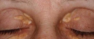
- cholesteatoma (tumor-like formation of dead epithelial cells and other substances surrounded by a capsule of connective tissue);
- perforation of the eardrum (large ruptures and extensive perforations);
- tympanosclerosis (overgrowth of connective tissue in the mucous membrane of the middle ear);
- disturbance in the chain of auditory ossicles;
- atelectasis of the tympanic cavity (strong retraction of the eardrum), etc.
Also, the cause of poor hearing and an indication for surgery may be congenital anomalies of the middle ear and mechanical damage to the eardrum (for example, due to careless removal of foreign objects from the ear).
The essence of the problem
When we talk about a form of surgery such as tympanoplasty (middle ear surgery), we mean a specific method for treating chronic purulent otitis media in the area of the eardrum.
It is important to note that such an effect is complex and has a significant restorative effect, allowing the reconstruction of the middle ear. It makes sense to pay attention to the fact that chronic otitis media is diagnosed quite often. It causes a hole to form in the eardrum, called a perforation. For this reason, it becomes possible for pathogenic bacteria to penetrate into the tympanic cavity, which, in turn, leads to the release of pus, which can bother you for many years.
https://www.youtube.com/watch?v=ytdev
Tympanoplasty is an operation without which it will be extremely difficult to overcome purulent otitis media. In addition, an advanced disease of this type can result in the occurrence of an abscess and thrombosis of cerebral vessels, and in some cases, meningitis.
Examination and preparation for surgery
Otoscopy
To prescribe surgery (or identify contraindications), an examination of the affected ear is prescribed, including:
- otoscopy – examination of the ear using special instruments;
- X-ray;
- bacteriological examination;
- audiometry - measuring hearing acuity using an audiometer (or tuning forks);
- passing all necessary tests.
In the preoperative period, the doctor prescribes general restorative therapy, medicinal sanitation (cleaning) of the middle ear, and other measures to improve ventilation and auditory function of the Eustachian tube.
Rehabilitation after surgery
After the operation, the patient is in the hospital for 1 to 3 days. Antibiotics are prescribed for 7–10 days. Anemization is carried out daily - the introduction of vasoconstrictor drugs into the pharyngeal mouth of the auditory tube. The ear canal is freed from tampons gradually, so the patient must come for examinations some time after discharge.
In order to feel comfortable in the postoperative period, it is necessary to avoid blowing your nose and sneezing, getting water into the ear (it is better to consult your doctor about how to wash your hair and shower), air travel, swimming pools and diving, as well as lifting large bodies. heavy objects.
Types of tympanoplasty
There are several types of such operations (classification according to G. Wulshtein), the choice of one of which is influenced by the nature of the damage to the ear structures:
Hearing restoration surgery
- Type I The indication for such an operation is the presence of perforation in the eardrum, but the chain of auditory ossicles must not be damaged and the patency of the auditory (Eustachian) tube must be maintained. This is the most common and simplest type of surgery. The defect is eliminated using the body's own tissues. To do this, the eardrum is lifted, a skin flap is placed over the hole and fixed on both sides using a special absorbable material. The effectiveness of this operation is about 95%. If necessary, repeated intervention may be performed;
- II type. This type of tympanoplasty is indicated for damage to such parts of the auditory ossicles of the middle ear as the manubrium, neck or head of the malleus, but the incus-stapedial joint must be intact. Plastic surgery involves prosthetics of damaged parts of the auditory ossicles;
- III type. Such plastic surgery is used in the absence of the eardrum, malleus and incus, but the stapes must be preserved. This operation does not involve the use of prostheses. Bone tissue from the body is used to restore the structures of the middle ear;
- IV type. It is advisable to perform such plastic surgery if all the auditory ossicles are missing, but the base of the stapes is preserved. In this case, the cochlear window is shielded using the preserved part of the eardrum or using a free flap;
- V type It is carried out in the case of a complete absence of all sound-conducting elements and involves fenestration of the semicircular canal (creation of a new opening in the bony labyrinth of the inner ear). Today, this type of plastic surgery is rarely performed; it has been almost completely replaced by stapedectomy (removal of the stapes and its replacement with a prosthesis).
If it is impossible to restore the eardrum and auditory ossicles at the same time, the operation is carried out in stages. First, reconstruction of the auditory ossicles (ossiculoplasty), and then plastic surgery of the eardrum (myringoplasty).
Depending on the type of operation, either local anesthesia or general anesthesia is used.
Diagnosis of ear malformations
To diagnose ear malformations, clinical and audiometric examinations, as well as radiological methods, are used. Accurate anatomical description of malformations using imaging techniques is indispensable, especially for planning, outcome of surgical middle ear reconstructions and cochlear implantation (CI).
Clinical examinations
Newborns with ear deformities should undergo a detailed examination of the craniofacial structures. A thorough examination of the skull, face and neck is necessary for configuration, symmetry, facial proportions, masticatory apparatus, occlusion, hair and skin condition, sensory functions, speech, voice and swallowing. The function of the middle ear is studied especially carefully, since the development of the outer ear closely correlates with the development of the middle ear. Parotid fistulas or appendages, as well as facial nerve paresis/palsy may accompany ear abnormalities.
In addition to the basic examination of the ears (inspection, palpation, photographic documentation), attention is paid to any anatomical features that may increase the risk or jeopardize the success of middle ear surgery. These features include dysfunction of the auditory tube due to hypertrophy of the adenoids, severe curvature of the nasal septum, as well as the presence of a cleft palate (and submucosa).
Ear deficiencies may be associated with syndromes; therefore, changes in the internal organs (eg, heart and kidneys), nervous system, and skeleton (eg, cervical spine) must be excluded by a multidisciplinary team that includes a pediatrician, neurologist, ophthalmologist, and orthopedist. Preoperative assessment of facial nerve function is mandatory if reconstructive middle ear surgery is planned.
Audiometry
Audiometry is the most important functional test in patients with ear problems. Severe defects of the external ear, such as congenital aural atresia, often occur in combination with severe disorders of the middle ear and can affect all its structures. In such cases, a conductive hearing loss of 45-60 dB occurs, and complete conductive blockade can often be found down to a level of approximately 60 dB.
In cases of unilateral congenital aural atresia, early hearing testing in the apparently normal contralateral ear is important to identify and rule out bilateral hearing loss. Depending on the degree of functional impairment, bilateral hearing loss can significantly interfere with speech development. Thus, early rehabilitation is mandatory (hearing aids first, surgical correction if necessary).
Read also: Severe cough in an adult. How to treat, relieve an attack.
Audiometry is possible even in an infant. Physiological studies include tympanometry (impedance measurements), otoacoustic emissions (OAE), and auditory evoked potentials (auditory brainstem responses). To establish specific threshold values, children around 3 years of age are subject to reflex and behavioral audiometric examinations, tympanometry, measurement of OAE and auditory brainstem response.
Objective measurement methods (OAE and auditory brainstem response) provide reliable results. In older children, reproducible results can be achieved by testing the auditory response using traditional pure-tone audiometry or behavioral audiometry. For accuracy, the audiological examination is repeated, especially in young children and in patients with multiple defects.
Vestibulological examination has differential diagnostic value. Violation of vestibular function does not exclude the presence of hearing.
Visualization methods
Traditional radiography is of little value in diagnosing ear defects. High-resolution computer tomography (HRCT), with its clear depiction of bony structures, is adequate to show changes in the external ear, external auditory canal (EAC), middle ear, and mastoid process. Magnetic resonance imaging (MRI) is best for imaging the membranous labyrinth, the neural structures of the internal auditory canal, and the cerebellopontine angle. HRCT and MRI are used in combination. Ultrasound diagnosis has no value for ear defects.
- CT scan.
HRCT of the temporal bone using a bone algorithm and slice thicknesses of 0.5–1 mm is suitable for assessing middle ear deficiencies. The traditional view is the axial plane, which shows both temporal bones and allows comparison of the two sides. Coronary scans are a useful important addition. Helical scanning technologies provide high spatial resolution without loss of quality and make it possible to document anatomical structures, visible variations and congenital or acquired deformities. Modern CT scans allow the reconstruction of secondary sections at any desired level in any plane, as well as the creation of three-dimensional structures.
HRCT visualizes the extent of the pneumatic cell system and the location of the jugular bulb, sigmoid sinus, and internal carotid artery. Moreover, HRCT shows the chain of auditory ossicles, the course of the tympanic and mastoid segments of the facial nerve, as well as the width of the internal auditory canal. The clear contrast between bone and air, as well as the high spatial resolution, makes this diagnostic procedure ideal for the middle ear.
Fixation of the ossicular chain cannot always be detected using HRCT. This may sometimes explain normal CT scans in patients with conductive hearing loss. The stapes is not always identified due to its small size; only sections of no more than 0.5 mm can show the entire stapes. Bone thickness of the cranial vault is often measured, particularly in the temporal and parietal regions, in patients who are scheduled to receive a bone-anchored hearing aid (BAHA). It is also possible to establish certain anatomical features when planning a CI.
Thus, HRCT not only indicates suitability for surgery, but also clearly indicates contraindications. Patients with a very atypical course of the facial nerve in the middle ear or severe middle ear disorders are not considered for surgical treatment.
- Magnetic resonance imaging.
MRI provides higher resolution than HRCT. Soft tissues are reflected in detail with the introduction of a contrast agent (gadolinium-DTPA) and using various sequences. MRI is unsurpassed in imaging fine detail in the temporal bone.
The disadvantage is the long examination time (about 20 minutes). The sections should be very thin (0.7-0.8 mm). T2-enhanced images (3D CISS sequences) are suitable for detailed imaging of the labyrinth and internal auditory canal. For example, cerebrospinal fluid and endolymph give a very strong signal, but nerve structures (facial nerve, vestibulocochlear nerve) give a very weak signal. MRI provides excellent data on the size and shape of the helix, vestibule, and semicircular canals, as well as the fluid content of the helix. Fibrous obliteration of the labyrinth can be detected using gradient images. The endolymphatic duct and sac can be seen and their size determined.
MRI is the only method for demonstrating the vestibulocochlear nerve at the same time as assessing the intracranial segments of the facial nerve. Therefore, this examination is necessary when planning a CI.
Genetic analysis
Ear deformities may occur in association with genetic syndromes. Therefore, patients with clinical suspicion of the syndromes should undergo molecular genetic testing. Genetic testing of patients' parents for an autosomal recessive or X-linked recessive disorder (heterozygous testing) is also recommended.
DNA mutations can be detected by laboratory analysis of blood samples. Healthy family members without clinical signs are tested for mutations to determine the likelihood of the disease. In the case of a family history of defects, intrauterine testing is performed.
In addition, genetic analysis is relevant in the differential diagnosis of hereditary diseases (including ear defects). Molecular genetic analysis is only meaningful when the causative genes are known and when the diagnosis leads to therapeutic consequences. In the context of molecular genetic testing, there should be detailed patient counseling.
Possible complications
Infection may cause pain
Possible complications after tympanoplasty include:
- infection accompanied by pain, swelling, discharge from the ear;
- no improvement in hearing;
- damage to bones and nerves, leading to various pathologies;
- displacement, atrophy or necrosis of the graft (if used);
- dizziness, imbalance;
- short-term paralysis of half the face.
In 3% of cases after tympanoplasty, hearing continues to decline until it is completely lost. Most often this occurs after non-radical tympanoplasty or complicated surgery.
Research to prepare for surgery
Before the actual surgical intervention, it is necessary to carry out a set of procedures to prepare the patient for the operation. This complex includes:
- Complete blood count (CBC/DIFF) with leukocyte formula/ESCBC/DIFF) with leukocyte formula/ESR;
- Blood type, Rh factor;
- Blood chemistry
Find out more
Seeing a doctor in a timely manner will help maintain your health.
Don't delay treatment, call right now. We work around the clock. tel. (24 hours a day)
Contraindications
Like any surgical intervention, tympanoplasty has contraindications. This operation cannot be performed if:
- exacerbation of chronic inflammatory processes in the middle ear;
- damage to the inner ear (labyrinth);
- intracranial or septicopyemic complications of ear diseases;
- significant damage to sound-conducting structures;
- impaired patency of the Eustachian (auditory) tube;
- general severe conditions.
If the second ear cannot hear at all, then the possibility of tympanoplasty is discussed individually, and the pros and cons are weighed.
Surgical intervention
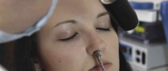
Almost all surgical interventions on the middle and inner ear are performed under general endotracheal anesthesia. On the day of surgery, the patient undergoes premedication, after which he is taken to the operating room on a gurney. It should be noted that the day before, if necessary, the hair in the area behind the ear is cut and shaved. Since one of the symptoms of labyrinthitis is a constant urge to vomit, such patients have to refuse food in the evening and morning before the intervention (so that complications do not arise during ear surgery). If the doctor will deal exclusively with the outer ear, it is permissible to perform surgery under local anesthesia.
Before surgery to restore hearing
- formation of a marginal or central perforation of the eardrum, which causes displacement of the auditory ossicles;
- polyps developing in the middle ear;
- tympanosclerosis;
- perforation of the eardrum, not accompanied by inflammatory processes;
- fibrosis of the middle ear.
Obviously, there are many reasons why surgery may be appropriate.
- exacerbation of chronic diseases;
- adhesive otitis media;
- complete deafness;
- severe general condition of the patient;
- sepsis and purulent complications.
Sanitation surgery on the middle ear with tympanoplasty is also contraindicated in cases where there is persistent obstruction of the auditory tube. This category includes congenital anomalies, as well as scars, as well as adhesions resulting from inflammation.
- acute inflammatory process in the middle ear;
https://www.youtube.com/watch?v=channelUCu_sa2ZIHKkr5IXqUe7_VpQ
— allergic diseases in the acute stage;
- problems with the upper respiratory tract;
- process of epidermization.
Only taking into account all the contraindications described above can a type of treatment such as tympanoplasty be prescribed. Reviews after surgery are an important sign of the quality of treatment, so it is always worth asking what patients think about a particular clinic. Forums can help with this.
It is worth remembering that the doctor is obliged to examine the other ear and make predictions regarding the risk of damage to it. After this, a decision will be made regarding surgery on the second eardrum.
A full-fledged general examination should also be carried out: coagulation test, biochemical and general analysis of blood and urine, as well as a blood test for syphilis, HIV, hepatitis B and C. The examination program also includes an ECG.
Tympanoplasty surgery is required in case of damage to the hearing aid, hearing loss of varying degrees, or the development of inflammation in the patient. The main indications for tympanoplasty are considered to be inflammatory processes in the patient that are chronic in nature, hearing loss, adhesive otitis media, tympanosclerosis, choleastomy and other diseases of the patient’s auditory system.
In preparation for tympanoplasty surgery, the patient must undergo the following measures: an appointment with an otolaryngologist (to rule out an ear infection), a thorough hearing test and a CT scan of the patient. In addition, the patient may need to undergo a number of tests to examine the hearing aid, undergo an electrocardiogram, and undergo the necessary tests for the patient (general tests, biochemistry).
Tympanoplasty can be performed both under local anesthesia and general anesthesia. The decision on the anesthesia used is made by the surgeon performing the operation and the anesthesiologist. The surgeon carries out the manipulations necessary to correct the defects through the auditory canal, thus preserving the appearance of the external organs of the hearing system.
On average, tympanoplasty surgery takes about 40-60 minutes. After the procedure is completed, a special drainage tube is installed in the wound. Its task is to collect discharge from the wound, preventing it from accumulating in the patient’s ear. Thus, the wound after surgery will heal faster. After 14-20 days, the drainage tube is removed.
During the operation, the material for the graft is skin taken from the non-hairy part of the scalp. The tympanoplasty operation makes it possible to get rid of pathological changes in the mucous membrane, remove carious bones, adhesions and cords. The formation of the tympanic cavity is usually accompanied by stopping the flow of suppuration and removing inflammation.
Tympanoplasty surgery plays a significant role not only in the treatment of patient diseases associated with hearing impairment, but is also important for the prevention of serious diseases in the future. After the operation, the healing process is accelerated, which helps the patient to recover quickly.
Depending on how the operation went, the patient remains in the clinic from several hours to a day, the doctor makes the appropriate decision. The postoperative period after tympanoplasty takes some time, and it may take 2-4 weeks before the patient’s final hearing recovery after surgery. During this time, the patient is strictly forbidden to blow his nose or allow water to get into his ear; he should try to avoid serious physical exertion, avoid visiting swimming pools, and air travel.
After tympanoplasty surgery, some complications are likely to occur. If the patient has identified a pathological condition or felt discomfort, the cause of which is presumably due to ear tympanoplasty, you should immediately consult a doctor, preferably the one who performed the operation.
Possible complications may include the appearance of signs of infection (development of fever or chills), swelling. Another alarm bell is discomfort that does not go away even after taking antibiotics. It is important to stop such complications at the initial stage so that they do not cause other serious problems.
The process of restoring the eardrum after surgery requires attention from the doctor and the patient. In the postoperative period after tympanoplasty, precautions must be taken. The main thing is to prevent liquids from getting into the ear, as this is often the cause of chronic otitis media.
Hearing loss
The group of complications includes the manifestation of hearing loss in the operated ear, which can reach the level of complete deafness. This problem makes itself felt after complicated or non-radical tympanoplasty. Impaired sound perception is sometimes a consequence of reactive labyrinthitis, which can resolve without treatment. If this does not happen, then competent therapy under the supervision of a doctor will help neutralize this problem.
Thus, you need to understand that hearing impairment caused by labyrinthitis is temporary and treatable.
But the good news is that such complications occur in only 3% of patients who have had surgery. Tympanoplasty reviews are mostly positive, so you should not be afraid of this technique.