V-Line surgery – to make your face oval!
A massive, wide, heavy jaw looks impressive in men, but does not adorn women. An elongated, angular or square lower part of the face does not correspond to the generally accepted canons of femininity, according to which facial features should be graceful and chiseled.
On the contrary, triangular, oval and intermediate V-shaped face shapes (oval in the form of a “heart”) have been considered a sign of aristocratic origin for centuries. The V-line of the jaw gives a soft, youthful charm to the features, since anatomically the lower third of a child's face is triangular in shape, which is the structure we associate with youth.
Types of surgery
Depending on the degree of organ damage, the doctor determines what type of intervention to perform. Medical practice distinguishes the following types of resection:
- Segmental, with a violation of the continuity of the jaw. It is carried out if the tumor begins to grow into the bone. With this diagnosis, radiation therapy rarely helps, so segmental resection is performed. The operation involves violating the integrity of the lower part of the face, which is fraught with serious cosmetic defects.
- Segmental, without breaking the continuity of the jaw. Another type of resection in which the integrity is not affected, since the operation is performed on a specific area and does not require complete removal of the lower part of the face. It is rarely performed, as it is prescribed in cases of secondary cancer, when the tumor affects the bone zonally.
- Segmental with disarticulation. It is performed when a patient is diagnosed with tumor growth inside the neurovascular bundle. Disarticulation involves isolating part of the jaw along the joint line without filing the bone. In this case, the bundle is carefully cut out along with the tendon of the temporal muscle.
- Half with disarticulation. The operation is indicated for marginal lesions of the corner of the mouth.
- Resection with soft tissues. It is carried out when working to remove areas located on the frontal area of the lower part of the face.
When choosing a surgical method, the surgeon focuses on the extent of tumor spread. When the tumor has grown on neighboring organs: tonsils, tongue, salivary glands and skin of the chin, these areas also need to be examined for the presence of harmful cells. After diagnosis, the doctor chooses which method will be optimal in the current picture of the disease.
Ideal proportions of the lower third of the face
The lower jaw consists of a horseshoe-shaped body and ascending branches, which are directed towards the temples and are located at an angle to the body of the jaw. If this angle exceeds 125°, the lower third of the face has an oval or triangular shape; with a less flat angle, the jaw looks square or round. The angle of the lower jaw is not only an anatomical, but also an age-related feature: in young children it is 150-160°, and the figure decreases with age.
The ideal proportions of the lower part of the face were calculated back in the Renaissance: a horizontal line is drawn from the middle of the interval between the lips and chin; when it intersects with the ascending branch of the lower jaw, it should form an angle of 100-110°. Such proportions create a “v-line”, that is, when viewed from the front, the line of the lower jaw resembles the Latin letter “V”.
Diagnosis and treatment of patients with deformities of the lower jaw in the angular area
The problem of diagnosing disproportion in the lower zone of the face and treating patients with this pathology, which is expressed in the form of a protruding angle of the lower jaw and causes an aesthetic problem for many people, especially females, is considered. A set of diagnostic methods is presented, which makes it possible to differentiate types of pathology into groups to facilitate the choice of treatment methods. Treatment methods that allow obtaining high aesthetic and functional results are presented.
The increase in the number of patients turning to a surgeon to change their face shape is due to increased migration, as well as the desire of women to have more graceful, sophisticated and at the same time proportional facial contours.
Progress in maxillofacial surgery has made it possible to consider the issues of diagnosis, planning and treatment of patients with deformities of the facial skeleton from different angles. However, a trend has emerged that cannot be ignored due to the growth of urbanization and the expansion of plastic surgery capabilities. A number of deformations have been identified, the treatment of which is aimed, rather, at the aesthetic aspects of facial anatomy, rather than at functional disorders of the dentofacial system. These are the so-called facial disproportions, which are characterized by deviations from the norm in the sizes of the lower, middle and upper zones of the face, both in the sagittal plane and in the transversal and vertical.
One of these deformations is the protruding angles of the lower jaw, which, according to the pathogenetic mechanisms of development, are related to hypertrophy of the masticatory muscles.
L. Whitaker first reported the possibility of reducing the width of the lower third of the face using osteotomy of the outer cortical plate of the angle of the mandible along with the masseter muscle. After Legg's first report of m. hypertrophy. masseter, many authors began to use partial myectomy m. masseter W. Adams and J. Converse proposed correcting this pathology surgically, using resection of m. masseter, and ostectomy of the angles of the lower jaw either externally or intraorally. Yang and Park, S. Baek et al. also proposed surgical classification and treatment, and divided patients into three groups, followed by surgical correction of the angular contours only. But the protruding angles of the lower zone of the face must be considered as a whole, from the point of view of the harmony of the entire face, and correction of not only the angles of the jaw, but also muscle hypertrophy, deformation of the chin, using a differentiated approach.
After analyzing foreign literature, we came across a huge variety of terms that can be used to designate disproportion in the lower zone of the face, characterized by wide and enlarged angles of the lower jaw. In the international classification, the term “hypertrophy of the masticatory muscles” is used, which characterizes changes in the bone tissue of the lower jaw. Since most of such work was carried out by scientists from Asia, where resection of the angles of the lower jaw is a popular operation, we decided to use their terminology, namely to call such a disproportion the protruding angles of the lower jaw, since this term most accurately characterizes it and we do not use this term in the domestic literature met.
Without dwelling on the description of the anatomy of the angle of the lower jaw, the parotid-masticatory region, which is well covered in the medical literature, it should be noted that manifestations of disproportion are caused by both the increase in the angle of the lower jaw (usually against the background of underdevelopment of the chin of the lower jaw), and by a change in the shape of the angle in the transversal and sagittal planes.
Most publications describe various methods for treating protruding mandibular angles. However, the authors did not conduct a thorough scientific study of diagnostic methods, did not differentiate the diverse forms of this pathology and did not determine the exact indications for the use of certain treatment methods, which was the reason for discussing this urgent problem of reconstructive plastic surgery.
facial surgery.
Material and methods
Over the past 10 years, 30 patients have come to us with protruding angles of the lower jaw. We spent at
They provide surgical and non-surgical correction of facial contours in order to improve its aesthetics and solve the psychosocial problems of patients. The age of the patients ranged from 19 to 42 years, there were 40 women and 6 men.
Preoperative diagnostics included the following examination methods: clinical examination, anthropometry, clinical photography, multislice computed tomography (MSCT), electromyography (EMG) of the masticatory muscles, ultrasound of the masticatory muscles themselves.
Clinical photometry was carried out using a semi-professional Nikon D500 SLR camera (focal length 1.5 m). Patients were photographed in frontal, profile, semi-profile and axial views. Particularly valuable results were obtained after 3D processing of data obtained using a multislice computed tomograph (Fig. 1).
Rice. 1. Three-dimensional construction of the facial skull.
This provided objective visualization, volumetric-spatial representation of the intervention area and made it possible to assess the volume of the upcoming correction. We obtained digital computed tomography data using MSCT performed on a SIEMENS Sensation 16 device. They were used to construct a three-dimensional model of the facial skull in the preoperative and postoperative periods, as well as to plan the possible results of the upcoming treatment.
Functional studies of the masticatory muscles are also important. Many of the patients who came to us with hypertrophy of the masticatory muscles themselves complained that, especially in the afternoon and at night, they noticed discomfort, expressed in tension and fatigue in the area of the angles of the lower jaw, as with one of the symptoms of bruxism. Patients suffering from bruxism identified by neurological examination were excluded from the study.
All patients with protruding mandibular angles underwent EMG recording the force of jaw compression. EMG was performed using the SINAPSIS device in the preoperative period, as well as in the early and late stages of the postoperative period. EMG made it possible to record and evaluate bioelectrical processes in the masticatory muscles, correlate the recorded values with the values obtained during a clinical examination, with MSCT and ultrasound data, and also outline a treatment plan. In addition, studying the electrical biopotentials of muscles helps to prevent such complications as weakness of chewing functions in the postoperative period, i.e. EMG data of the masticatory muscles objectively show the maximum volume of myectomy.
The above examination methods were used in the preoperative, postoperative periods and in the long term after treatment. Analysis of data from all examination methods obtained at the preoperative stage allowed us to classify patients based on the morphological features of the deformities:
Group 1 - mild deformity with protrusion of the corners in the posteroinferior direction, i.e. in the sagittal plane, which reveals the facial profile;
2nd group - noticeable protrusion of the corners not only in the sagittal plane, but also in the lateral (main deformity);
3rd - there are pronounced features of the 2nd group in combination with hypertrophy of m. masseter; 4th group - the deformation is based only on hypertrophy of m. masseter
Of our 30 patients, 6 were assigned to group 1, 11 to group 2, 9 to group 3, and 4 to group 4.
Patients of the first 3 groups were treated surgically, patients of the 4th group received botulinum toxin injections
type A in a dose of 20 units. into the chewing muscles themselves.
Operation techniques
All operations were performed under general anesthesia with endotracheal intubation. We only used
intraoral surgical access. The incision of the mucous membrane began in the retromolar region 1 cm above the last lower molar and then continued along the lower buccal-alveolar fold of the vestibule of the mouth to the projection of the 1st lower molar. Then you should go to the periosteum, covering the area of the bone of the lower jaw behind and downward from the last lower molar. The periosteum is dissected and exfoliated along with the fibers attached to it m. masseter to the area required to open the entire angle of the lower jaw. In patients of the 1st group, marginal resection of the mandibular angle with bending osteotomy was then used, in patients of the 2nd group - sagittal splitting ostectomy of the mandibular angle, in patients of the 3rd group - sagittal splitting ostectomy of the angle and partial resection of the masticatory muscles themselves (Fig. 2) .
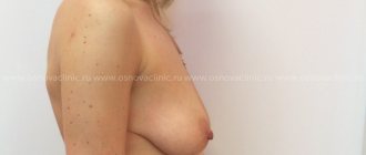
Rice. 2. Patient A. before (left) and 6 years after surgery (right).
During myectomy, the internal fibers of m. were resected along the sagittal plane. masseter The volume of bone and muscle resection was determined according to data obtained after calculations and processing of the results of the preoperative examination, and was agreed with the patient. After removing the excess mandibular angle, the so-called contouring of the angles was carried out, as a result of which it was possible to obtain the maximum natural shape of the mandibular angle (Fig. 3, 4).
Rice. 3. Patient E. before surgery.
Rice. 4. Patient E. after surgery.
In the early surgical period (days 15–20) and in the long term after surgery (1–5 years), we used the same examination methods as before treatment.
According to an analysis of the results of treatment of patients with protruding angles of the lower jaw: in 25 out of 30
the person achieved a satisfactory result, the square contours of the lower area of the face were corrected
more refined, the face became proportional, acquired an oval shape, and symmetry was preserved. In 1 patient, asymmetry persisted for 1 month, associated with hematoma and asymmetrical edema that occurred in the early postoperative period. After 1 month, the asymmetry gradually disappeared spontaneously; no special re-surgical correction was required. All patients in group 4 achieved partial correction; the effect lasted an average of 3-4 months, after which additional botulinum toxin injections were required (Fig. 5).
Rice. 5. Patient R. before (left) and after (right) treatment with botulinum toxin.
Conclusion
The results of studies conducted in 30 patients with protruding angles of the lower jaw, the complex of diagnostic methods we developed and the distribution of patients into clinical groups depending on anatomical and anthropometric indicators turned out to be effective, which contributed to obtaining good aesthetic and functional results.
Authors:
Doctor of Medical Sciences F.H. Nabiev, asp. K.V. Filippov, asp. P.V. Libin
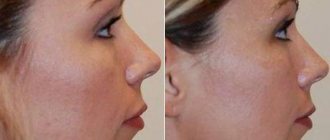
Reshaping the lower jaw with V-Line surgery
The possibilities of modern surgery make it possible to change the shape of the lower third of the face and bring it closer to the desired oval or triangle.
Depending on the initial shape of the lower jaw, V-Line plastic surgery (Jaw Reduction, V-Line Mandibular Contouring) may include:
- changing the contour of the lower jaw by resection of the corners
- straightening the edge of the jaw body using grinding
- narrowing of the chin using T-osteotomy (excision of a small section of bone tissue in the center or pushing it down and forward)
Mandibuloplasty is performed under general anesthesia and lasts an average of 1.5 hours. In the vast majority of cases, intraoral access is used in the retromolar region (behind the last teeth) and along the lower arch of the vestibule of the oral cavity (under the lower front teeth), which does not leave marks on the face. The period of postoperative hospital stay is 1 night.
The active rehabilitation period lasts 10-14 days: during this period it is necessary to use a face mask, there are some restrictions on food. The face recovers completely after surgery within 2 months.
V-Line bone grafting does not disrupt the structure of the dentition and is easily tolerated. The operation is indicated for patients aged 20-25 years with fully formed bone tissue.
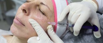
Contour plastic of the lower jaw
Mandibular contouring is a cosmetic procedure for correcting the relief of the lower jaw line, chin and bitter folds, based on the injection of natural fillers based on hyaluronic acid into problem areas.
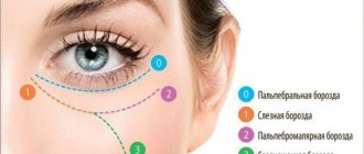
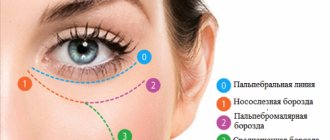
The chin is an unpaired area of the middle part of the lower jaw, bounded above by the chin-labial groove, below by the base of the lower jaw, on the sides by conventionally drawn lines from the corners of the mouth vertically downwards. The individual shape of each person’s chin is determined by the anatomical features of the bone structures (relief of the lower jaw, mental protuberance), the muscles of this area, and the deep and superficial layers of subcutaneous fat. The mental tubercle is the protruding part of the mental region of the mandibular bone, located along its lower edge. The most common reason for patients to come to us is a “weak” or sunken chin and a strongly protruding chin. Within this problem, contour plastic surgery is an additional procedure after surgical correction. As an independent procedure, it is indicated for such problems as a “double chin” (a strongly pronounced transverse groove in the projection of the hyoid bone on the neck downward from the lower edge of the mandibular bone), age-related drooping of the soft tissues of the lower third of the face, loss of skin elasticity after sudden weight loss and, as a result, , formation of pronounced wrinkles and folds in the chin area.
The bitter fold is a groove that forms during the aging process from the corners of the mouth down to the lower edge of the lower jaw, limited by the depressor anguli oris muscle.
Let's look at the anatomy of the muscles of the chin region:
- The depressor anguli oris muscle is a thin flat paired muscle of a triangular shape, its base is attached to the oblique edge of the lower jaw on the sides of the chin, and its apex is woven into the fibers of the orbicularis oris muscle in the area of the corner of the mouth. This muscle is the border of the bitter fold;
- The depressor labii inferioris muscle is a thin flat diamond-shaped paired muscle located under the depressor anguli oris muscle; its upper edge is woven into the deep skin layer of the lower lip, extends laterally downwards and is attached to the outer oblique edge of the lower jaw. Together with the mentalis muscle, it forms the mental fold;
- mentalis muscle - a paired muscle running from the mental fossa obliquely down to the lower edge of the mental region;
- platysma - a flat quadrangular subcutaneous muscle of the neck, covering the anterior edge of the sternocleidomastoid muscle, attached to the lower edge of the mandibular bone and woven into the fibers of the muscle that depresses the angle of the oris;
- The orbicularis oris muscle is a developed muscle, the fibers of which are intertwined with all the muscles in this area.
All of the above muscles are actively involved in articulation, therefore, with age, they form numerous wrinkles, folds and furrows on the skin, the most expressive of which is the bitter fold.
Also, a significant role in age-related changes in the relief of the lower jaw line is played by the points of skin fixation at the site of the mandibular ligament and the branch of the facial nerve.
The appearance of drooping cheeks is also explained by the presence of the mandibular, mental and parotid septa. Being tightly attached to the underlying tissues, these septa remain motionless, while the surrounding tissues sag due to age and loss of elasticity.
Changing the shape and relief of the chin is usually carried out using a denser filler based on hyaluronic acid, sometimes calcium hydroxyapatite, and filling deep wrinkles with a less viscous filler.
Best results with 3D planning
The V-Line operation is designed to create a feminine and aristocratic oval face, an elegant small chin, and a more youthful appearance by changing the jaw line.
The VIP Studio virtual plastic laboratory makes it possible to 3D model the ideal proportions and desired shapes before surgery. Based on computed tomography and light scanning data, an accurate three-dimensional model of the face, head and neck is formed. The surgeon and the patient jointly plan the scope of the intervention; based on the model, calculations are made that are used during the operation.
Thanks to the VIP Studio virtual plastic technology, which Dr. Guryanov uses in his work, the result of the operation is as close as possible to the client’s expectations.
Technique
The purpose of the operation is to eliminate the tumor.
If cancer or sarcoma is detected, the doctor treats it as follows:
- the patient is placed on the surgical table on his back, and a cushion is placed under his shoulders,
- actions to administer anesthesia are performed,
- The lower lip is dissected along the midline, and then the tissue of the chin,
- after this, the incision is made along the edge of the affected bone and the back of the branch 5 cm above the angle of the lower jaw,
- if the treatment is accompanied by the removal of lymph nodes, then the dissection is performed from the chin to the mastoid process. Another 1 incision is made along the anterior part of the sternocleidomastoid muscle,
- in the area of the vestibule of the mouth, the mucous membrane is cut to the bone, and then at the same distance an incision is made in the oral mucosa,
- remove tissue starting from the chin,
- extract the tooth
- the inner part of the movable bone of the skull is sawed down along the midline, and then taken outward,
- cut with a scalpel the internal tissues of the body of the bone to be removed to the coronary branch,
- the medial pterygoid muscle is cut off with scissors,
- the incision is brought to the mandibular foramen, crossing the alveolar artery, which is subsequently ligated,
- use scissors (nippers) to remove the coronal branch,
- the affected part of the jaw is pulled down and turned inward to make an incision on the temporomandibular joint,
- dissect the lateral pterygoid muscle,
- perform displacement of the articular surface of the jaw from its cavity, simultaneously removing the affected tissue,
- self-absorbing surgical sutures are applied to close the exposed part of the bone,
- suturing the mucous membranes of the cheeks and oral cavity,
- turundas made of gauze are placed in the cut out part for several days to prevent the occurrence of a hematoma,
- Various types of splints are installed to prevent displacement of bone elements.
Benign tumors are not accompanied by dissection of the lower lip. In this case, the incision is made along the lower edge of the submandibular triangle so as not to damage the mandibular vein. The affected area is cut out in a special way. Lymph nodes cannot be removed. The resection is completed with the installation of a graft.
When performing the operation, it is necessary to follow all the rules of the procedure, since the mandibular nerve can be damaged, which will cause loss of sensitivity in the lower lip and chin.
After resection, antibacterial and anti-inflammatory therapy is prescribed. Suture removal is carried out on an individual basis and depending on the patient’s condition. Physical activity is also not recommended in the next 2 weeks.
How is plastic surgery of the angles of the lower jaw performed?
After planning the implant, it is time for surgery. It lasts about an hour under local anesthesia. We usually use intraoral access, so there are no stitches on the face after surgery.
Of course, jaw augmentation surgery, like any plastic surgery, requires time for rehabilitation. The recovery period lasts about two weeks, and for the first few days you will have to wear a bandage that helps stabilize the implant.
Subsequently, there is no danger of it migrating: the pores of the material from which the implant is made grow with connective tissue so that the implant simply grows into the bone tissue. The implant is installed once and for life, does not require replacement.
What a beautiful jaw a woman has
The structure of a woman's face has its own characteristics. In order to look beautiful and graceful, a woman needs chiseled cheekbones and a smooth jawline.
Despite the widespread dictate of Angelina Jolie's image, the convex angles of the lower jaw and protruding chin are a manifestation of masculine traits. A feminine jawline should be smoother, yet firm and well-defined.
To make the image refined and sophisticated, too pronounced angles of the lower jaw require delicate grinding of the bone tissue.
Jaw correction may be accompanied by chin augmentation and removal of Bisha's lumps to enhance the cheekbone line. These procedures allow you to tighten the skin under the chin, get rid of the double chin effect, and create a beautiful line of the chin-cervical angle.