What is erysipelas?
A skin disease that is infectious in nature is called erysipelas. The disease is caused by streptococcus.
Streptococci cause an infectious disease - streptoderma. Here you can read about the treatment of streptoderma in adults.
The infection penetrates through injured areas of the skin:
- Cracks.
- Wounds.
- Abrasions.
- Scratches.
Sometimes there are cases of infection through the endogenous route:
- airborne;
- through infected objects;
- food.
In this case, the spread of streptococcus passes through the human lymphatic system.
Provoking factors
The development of erysipelas on the face can be provoked by:
- stress loads;
- long-term fatigue;
- increased sports loads;
- sudden temperature changes;
- mechanical skin defects;
- Tan.
One of the obvious reasons may be:
- Reduced immunity.
- Chronic somatic diseases.
- Unbalanced diet.
- Alcohol abuse.
- Streptococcus infection.
What does a erysipelas look like on your face?
The disease “erysipelas” on the face is often expressed through visual signs:
- Disruption of lymph flow, as streptococcus affects the lymphatic vessels.
- Cracks appear.
- Soreness in inflamed areas.
- A red spot appears that rises above the level of the skin.
- Erysipelas on the face always have clearly defined boundaries.
- After some time, the spot grows and becomes hot.
- In advanced stages, blisters and hemorrhages may appear.
With erysipelas located in the face area, the inflammatory process can spread in the following areas:
- ears;
- nose;
- ear opening;
- mouth;
- cheeks;
- rarely located under the hairline.
In another article you can see what erysipelas on the leg looks like.
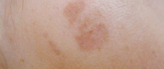
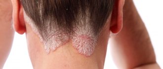
Photo
To determine the visual symptoms of erysipelas, use the provided photographic material.
Symptoms
The initial symptomatic manifestations of erysipelas are:
- headache and general weakness;
- temperature rise up to 40 degrees;
- aches in the joints, lower back, muscles;
- burning sensation;
- itching;
- gagging;
- swelling in the damaged area;
- tachycardia.
There may be pain in the scalp, often in inflamed areas.
In severe conditions the following are observed:
- vomit;
- convulsions;
- rave.
Clinical picture[edit | edit code]
The incubation period can only be established for post-traumatic erysipelas; in these cases it lasts from several hours to 3-5 days. In more than 90% of cases, erysipelas begins acutely; patients indicate not only the day, but also the hour of its occurrence.
General toxic syndrome precedes local changes. A rapid rise in temperature is accompanied by chills, often shaking. Severe signs of intoxication are revealed - headaches, dizziness, weakness, nausea, and possible vomiting. In severe cases, there may be seizures and delirium. Local symptoms appear 10-20 hours after the onset of the disease.
At first, patients experience itching in limited areas, a feeling of sweating, and tightness of the skin. Then swelling and pain appear in these places, corresponding to the development of regional lymphadenitis, sometimes with lymphostasis (on the inside of the thigh). The pain is most pronounced with erysipelas of the scalp. Quite often, pain occurs in the area of the regional lymph nodes, which intensifies with movement. Then redness of the skin (erythema) with swelling appears.
A spot of bright hyperemia with clear, uneven boundaries in the form of “tongues of flame” or “geographic map”, swelling, and thickening of the skin forms on the affected area. The lesion is hot and slightly painful to the touch. In case of lymph circulation disorders, hyperemia has a cyanotic tint; in case of trophic disorders of the dermis with lymphovenous insufficiency, it is brownish. After pressing with your fingers on the area of erythema, the redness underneath disappears within 1-2 seconds. Due to the stretching of the epidermis, the erythema is shiny, and at its edges the skin is slightly raised in the form of a peripheral infiltration ridge. At the same time, in most cases, especially with primary or recurrent erysipelas, the phenomena of regional lymphadenitis are observed: compaction of the lymph nodes, their pain on palpation, limited mobility. In many patients, concomitant lymphangitis appears in the form of a narrow pale pink stripe on the skin connecting the erythema with the regional group of lymph nodes.
From the internal organs, muffled heart sounds, tachycardia, and arterial hypotension can be observed. In rare cases, meningeal symptoms appear.
After the start of treatment, fever, varying in height and nature of the temperature curve, and other manifestations of toxicosis usually persist for 5-7 days, and sometimes a little longer. As body temperature decreases, a period of convalescence begins. The reverse development of local inflammatory reactions occurs after body temperature normalizes: the erythema turns pale, its boundaries become unclear, and the marginal infiltration ridge disappears. The swelling subsides, the symptoms of regional lymphadenitis decrease and disappear. After the hyperemia disappears, fine scaly peeling of the skin is observed. Local manifestations of erysipelas disappear by the 10th - 14th day of illness; pastiness and pigmentation of the skin may persist for a long time. In some cases, regional lymphadenitis and skin infiltration persist for a long time, which indicates the risk of early recurrence of erysipelas. Long-term persistence of persistent edema is a sign of the formation of lymphostasis. The given clinical characteristics are characteristic of erythematous erysipelas.
Without treatment, the process progresses rapidly, and so-called creeping or metastatic forms occur. In this case, complications of a septic nature arise. The appearance of blisters filled with serous-yellowish contents against the background of the plaque characterizes the development of the erythematous-bullous form of the disease. The size of the bubbles can range from very small to large drain ones. After the bubbles dry, dense crusts remain. The erythematous-hemorrhagic form is characterized by the appearance of pinpoint or more extensive hemorrhages. Bullous and especially hemorrhagic forms are characterized by a predominant severity of the course and often lead to persistent lymphostasis.
The clinical picture of early and late recurrent erysipelas is basically the same as that of primary erysipelas. A feature of the often recurrent form is the mild severity of the general toxic syndrome: temperature 37.5-38.5 ° C for 1-2 days with very moderate symptoms of intoxication, greasy erythema without edema, poorly demarcated from the surrounding skin. The occurrence of a recurrent form of the disease is facilitated by concomitant diseases (chronic venous insufficiency, lymphostasis, diabetes mellitus, chronic foci of streptococcal infection), as well as unfavorable professional conditions (the need to stand for many hours, hypothermia) and old age.
Forms of erysipelas
There are several classifications of erysipelas.
Depending on the frequency of occurrence, there are varieties:
- Primary. The patient is diagnosed with the disease for the first time. It most often affects the face area.
- Repeated erysipelas. Occurs repeatedly, localization can be varied.
- Recurrent. The manifestation of the disease can be early or late. Localize both in new zones and meet in old places.
According to the degree of progression, erysipelas is divided into the following stages:
- Easy stage. It is short-term, its development takes from 1 to 3 days. Characterized by:
- high body temperature;
- moderate intoxication;
- erythematous skin lesions.
- Medium-heavy. It also occurs quite quickly; this stage takes up to 5 days. Expressed:
- high body temperature;
- general weakness;
- severe headaches;
- has extensive areas of inflammation.
- Heavy. The longest stage of the erysipelas process on the face. Often this stage is complicated by infectious lesions:
- sepsis;
- pneumonia;
- infectious-toxic shock.
There are also several forms of erysipelas depending on the nature of the local manifestation:
- Erythematous erysipelas. With this form of erysipelas, symptoms are observed: the affected area has redness, swelling is observed, and the affected area is painful. Visually this form can be distinguished:
- Has a bright color.
- Clear outlines.
- The inflammation can grow and rise above the skin level.
- The edges take on an irregular shape.
- Peeling of the skin appears.
- Erythematous bullous erysipelas. This form has similar symptomatic features. However, after 3 days the following symptoms are observed:
- detachment of the upper layer of the skin occurs;
- bubbles with transparent contents form;
- after the formations burst, a brown crusted formation appears;
- in the area of the former vesicles, erosions are observed, which, without treatment, develop into trophic ulcers;
- There are phenomena of intoxication and lymphadenitis.
- Erythematous-hemorrhagic erysipelas. This form of erysipelas is expressed through hemorrhages in the area of the inflammatory process. Symptomatic signs added:
- hemorrhages;
- duration up to two weeks;
- fever;
- the development of necrotic lesions on the skin is possible.
- Bullous-hemorrhagic erysipelas. A distinctive feature of this form of erysipelas and erythematous-bullous is the formation of bloody filler inside the blisters. Refers to the most severe forms of erysipelas. Symptomatic manifestations include:
- vascular damage;
- there is serous-hemorrhagic content inside the vesicles;
- after the blisters open, erosions may form;
- the blisters can leave behind scars.
You might be interested! Demodicosis in women: how to recognize and how to treat?
There is a classification of erysipelas and inflammatory processes according to the distribution of forms of local manifestation:
- Localized. The source of spread does not extend beyond one zone. For example: face, legs or back.
- Common. Erysipelas are observed in several places at the same time.
- Migratory. In one place the pathological process may subside, while in another there is an increased development of inflammation.
- Metastatic. Pathological areas may be located far from each other.
Classification[edit | edit code]
The modern clinical classification of erysipelas includes the following forms of the disease.
- By the nature of local lesions:
- erythematous;
- erythematous-bullous;
- erythematous-hemorrhagic;
- bullous-hemorrhagic.
- According to the degree of intoxication (severity):
- light;
- moderate severity;
- heavy.
- By flow rate:
- primary;
- repeated;
- recurrent (often and rarely, early and late).
- According to the prevalence of local manifestations:
- localized;
- widespread;
- wandering (creeping, migrating);
- metastatic.
Treatment
Complications observed with erysipelas:
- rheumatism;
- sepsis;
- nephritis;
- myocarditis;
- abscess;
- elephantiasis of the limbs.
Therefore, timely diagnosis and treatment of this skin lesion is necessary.
Diagnostic criteria for identifying erysipelas are:
- Acute onset of the disease.
- The severity of symptomatic intoxication.
- Erysipelas mainly spreads in the face area, less commonly in the limbs.
- Local lymphadenitis may develop.
- Development of erythema.
- Absence of pain in the affected area during periods of rest.
Very often, erysipelas has similar symptomatic manifestations to:
- abscess;
- phlegmon;
- erythema nodosum;
- eczema;
- herpes zoster;
- thrombophlebitis.
Drug treatment.
The causative agent of erysipelas is hemolytic streptococcus.
To combat infection, antibacterial drugs of various groups are used:
- penicillins;
- sulfonamides;
- nitrofurans.
Prescribed medications such as:
- oleandomycin;
- erythromycin;
- ampicillin trihydrate;
- clindamycin.
Various combinations of dosage forms may be prescribed.
The therapeutic effect is expressed as follows:
- body temperature decreases;
- inflammatory areas turn pale;
- skin cleansing occurs after 3 days.
Local medications are also prescribed:
- powders such as enteroseptol;
- ointments made from crushed tablet medications;
- erythromycin ointment.
Antihistamines and non-steroidal medications are also prescribed.
In severe forms, the following are possible:
- blood transfusion;
- administration of placental gamma globulin;
- vitamin complexes;
- biostimulants: levamisole, methyluracil, pentoxyl.
Physiotherapy
Physiotherapy is prescribed to reduce inflammation in pathological areas, as well as to reduce the severity of intoxication.
Such manipulations include:
- ultraviolet irradiation, which has an antibacterial effect;
- drug electrophoresis;
- UHF therapy, the use of electromagnetic waves on the affected areas. It is also used to get rid of boils on the body. Here you will find in more detail the answer to the question about the causes, treatment and photos of furunculosis.
- Microwave therapy, treatment of the zone with electromagnetic frequencies.
Surgery
When treating bullous forms, the use of local antiseptics does not have the desired effect, but can only increase exudation and inhibit the healing process.
When the disease develops, the following have a good effect:
- cryodestruction;
- quartzing;
- laser therapy.
Pathogens[edit | edit code]
Erysipelas is a common streptococcal soft tissue infection. Erysipelas can be caused by any serovar of group A beta-hemolytic streptococcus. Streptococci are relatively resistant to environmental conditions. A sporadic increase in incidence is observed in the summer-autumn period, the entrance gates of infection are minor injuries, abrasions, and abrasions.
The source of infection is a patient with any form of streptococcal infection or a streptococcal carrier. There is a special selective susceptibility or predisposition to erysipelas. Some people get sick many times because the developing immunity is unstable. Streptococci enter the body through minor damage to the skin and mucous membranes. Exogenous infection is possible (contaminated instruments, dressings), as well as from chronic streptococcal foci of infection (for example, in patients with chronic tonsillitis). In this case, the state of reactivity of the body is of decisive importance, causing wide fluctuations in susceptibility to infectious pathogens, in particular to streptococci.
Prevention
Since erysipelas is a contagious disease, it is necessary to reduce or cancel all contacts with infected people. Family members should prevent injuries to the skin and monitor the sanitary and hygienic condition of the skin.
For preventive purposes, it is better to adhere to certain rules:
- promptly treat all painful conditions of the skin;
- observe personal hygiene rules, especially after visiting crowded places;
- do not allow infectious agents to get into small scratches or wounds;
- treat all violations of the integrity of the skin.
- timely treatment of fungal skin infections and lymphovenous insufficiency.
Differential diagnosis[edit | edit code]
Erysipelas is differentiated from many infectious, surgical, skin and internal diseases: erysipeloid, anthrax, abscess, phlegmon, felon, phlebitis and thrombophlebitis, obliterating endarteritis with trophic disorders, eczema, dermatitis, toxicoderma and other skin diseases, systemic lupus erythematosus, scleroderma, Lyme disease (borreliosis), etc.
When making a clinical diagnosis of erysipelas, take into account the acute onset of the disease with fever and other manifestations of intoxication, which often precede the occurrence of typical local phenomena (in some cases occurring simultaneously with them), the characteristic localization of local inflammatory reactions (lower limbs, face, less often other areas of the skin integument), development of regional lymphadenitis, absence of severe pain at rest.