Seroma of postoperative scar
Doctors often have to perform urgent and complex operations. Many of them literally save a person’s life. Sometimes it happens that after surgery complications arise in the suture area. After mechanical dissection of the skin, lymph sometimes accumulates in places where scars form. Between the suppression of capillaries and the fatty layer there is a large accumulation of serous fluid. When its volume increases greatly, the fluid begins to seep through the scar tissue, if it is not very dense.
This physiological phenomenon causes a lot of discomfort. The consistency of the liquid can be viscous or liquid. Usually its color is straw-yellow, sometimes gray with bloody streaks. It has no smell. The disease occurs with large swelling in the suture area, often with painful sensations.
Seroma often appears after plastic surgery. Most of these complications go away on their own within a couple of weeks. However, after problems with a large amount of fluid, the skin begins to sag a lot. All these moments cause discomfort and anxiety. As a result of such complications, the patient is forced to visit the doctor more than once and spend a long time recovering.
Such complications do not cause severe pain. Sometimes pain is felt if there is too much fluid. It is quite difficult to immediately recognize such a complication.
Main reasons
After the operation, various factors are noted that cause fluid to form. The main reason is a large wound surface and detachment of large surfaces of subcutaneous tissue. The wound surface is associated with damage to a large number of lymph vessels. They are not able to thrombose like blood vessels. This leads to the accumulation of serous fluid , which is lymph. Bloody discharge gives it a red color.
There are also other reasons that cause similar complications after surgery. These include:
- Traumatic work with tissues - during the operation, the surgeon must work gently with tissues. The cuts must be made carefully in one movement. Numerous incisions result in a large area of damaged tissue, which creates the risk of seroma formation.
- Inflammatory process - much in the postoperative period depends on the skin and the body. Sometimes the process of prolongation occurs with developing infectious inflammation. As a result, excess lymph fluid is formed.
- Excess weight - very often people with excess fat experience poor wound healing. This situation is observed in 75% of patients in this group.
- Tissue burn - in this case, necrosis occurs and an inflammatory fluid is formed. Coagulation during surgery must be applied with extreme precision.
- Diabetes mellitus - with this diagnosis, the concentration of glucose in the blood increases. It creates obstacles to the normal restoration of damaged tissues.
- Dense subcutaneous fat layer - if the thickness of the layer exceeds 5 cm, then you need to be prepared for the appearance of seroma. To avoid complications, it is necessary to undergo liposuction before surgery.
- Age - in elderly patients, all metabolic processes occur more slowly. As a result, new cells are not formed as quickly.
- Hypertension - lymph under high pressure in the body is distributed unevenly.
Treatment of seroma after surgery with folk remedies
Serous fluid is not the biggest postoperative problem, but some complications can still arise that cause discomfort to the person. Fluid accumulation occurs at the intersection of capillaries. That is, lymph accumulates within the cavity, which is located near the aponeurosis and fatty tissue under the human skin.
That is why such complications most often occur in dense people with a large layer of fat under the skin. During the development of a disease associated with serous fluid, a straw-colored discharge may appear, which does not have an unpleasant odor, but severe swelling may appear, and sometimes a person even feels pain at the site of seroma accumulation.
Most often, the accumulation of serous fluid occurs precisely after surgery. For example, we can distinguish plastic surgeries, after which fluid accumulates, which leads to negative consequences. These side effects do not affect human health in any way, but undesirable phenomena such as sagging skin in places where fluid accumulates may still appear, which of course spoils the aesthetic appearance of a person. In addition, seroma increases the healing time of the skin, and because of this you have to visit the doctor more often, which also causes inconvenience.
Theoretically, seroma can occur after any violation of the integrity of lymph vessels, which do not “know how” to thrombose quickly, as blood vessels do. While they are healing, lymph continues to move through them for some time, flowing from the rupture sites into the resulting cavity. According to the ICD 10 classification system, seroma of the postoperative suture does not have a separate code. It is assigned depending on the type of operation performed and the reason that influenced the development of this complication.
In practice, it most often occurs after such cardinal surgical interventions:
- abdominal plastic surgery;
- cesarean section (this postoperative suture seroma has ICD 10 code “O 86.0”, which means suppuration of the postoperative wound and/or infiltration in its area);
- mastectomy.
As you can see, it is mainly women who are at risk, and those who have solid subcutaneous fat deposits. Why is that? Because these deposits, when their integral structure is damaged, tend to peel off from the muscle layer. As a result, subcutaneous cavities are formed, in which fluid begins to collect from the lymph vessels torn during the operation.
The following patients are also at risk:
- those suffering from diabetes;
- elderly people (especially overweight);
- hypertensive patients.
The accumulation of serous fluid in the area where the surgical suture is located is caused by the presence of a wide variety of factors that took place at the time of surgery.
The main causes of seroma development are:
- Excessive activity of lymphatic capillaries. Even an operation that does not pose a threat to health is always local stress for the body and skin that has been injured by a mechanical cut. Under such conditions, lymphatic capillaries begin to actively synthesize lymph and redirect it to the surgical site. As a result of an abnormal reaction of the lymphatic system, the patient faces very unpleasant consequences.
- Inflammatory process. Each body reacts differently to surgery. Some people's skin and soft tissues recover quickly and without complications, while there are patients who develop non-infectious inflammation of the wound surface with excessive accumulation of lymphatic fluid.
- Hypertonic disease. High blood pressure can be a factor in the irrational distribution of lymph to all parts of the body.
- Overweight. At least 75% of all surgical patients who are overweight face the problem of postoperative suture healing and the accumulation of serous fluid. The presence of a large amount of fatty tissue contributes to this. Patients who have elastic muscles in the abdominal area almost never encounter the problem of seroma.
- Diabetes. This is a concomitant disease that is characterized by increased concentrations of glucose in the blood. Excess sugar does not allow blood vessels and the circulatory system as a whole to function normally and restore damaged tissue.
- Senile age. As you age, the intensity of metabolic processes in the body decreases. The division of epidermal cells, blood, soft tissues and the formation of lymph slows down. Therefore, deviations in the recovery process and the formation of serous fluid at the incision sites are possible.
Most of these potential causes that can cause postoperative complications are identified by doctors several days before surgery. The patient takes a blood test to check blood sugar levels, coagulability, and the presence of chronic diseases of infectious origin. A comprehensive examination of the body, all its organs and systems is also carried out. Therefore, if some pathology has been established, the patient is immediately prescribed specific treatment after the operation in order to prevent the development of seroma. For example, in a patient with diabetes mellitus, during the recovery period, insulin administration is increased to the maximum limit in order to lower the level of glucose concentration in the blood as much as possible and prevent tissue necrosis around the suture, as often happens in patients with this endocrine disease.
Seroma can be suspected if the following symptoms are present:
- The patient feels as if fluid is beginning to overflow in the lower abdomen.
- Sometimes there is swelling and a feeling of bulging in the lower abdomen. Patients claim that their abdomen has suddenly increased in volume, although this was not the case a few days ago.
If the serous fluid has reached large volumes , then the following symptoms occur:
- Soreness or a feeling of tension in the area where the seroma has accumulated. Most often this is the lower abdomen.
- Nagging pain that begins to intensify if the patient gets to his feet.
- Redness of the skin in the place where the seroma has accumulated the most.
- General weakness, increased body temperature up to 37 degrees, fatigue.
Diagnosis of seroma is based on examination and instrumental research methods.
- Inspection. During the examination, the surgeon will notice the presence of swelling in the lower abdomen. On palpation, fluid flows from one side to the other, a fluctuation indicating that there is an accumulation of fluid. In addition, the presence of seroma symptoms will leave no doubt for making the correct diagnosis.
- Instrumental research methods - ultrasound of the soft tissues of the abdomen. With ultrasound, the accumulation of fluid between the muscles of the anterior abdominal wall and subcutaneous fat is very clearly visible. Taking into account all the symptoms and ultrasound scan results, it does not seem difficult to diagnose seroma.
In most postoperative cases, seroma resolves within a few days. Throughout this period, the patient is observed by the surgeon and follows his recommendations for restoring the body. If fluid does accumulate and there is a risk of infection or blood infection, treatment will be needed.
Seroma is treated in two ways:
It is considered the easiest way to remove seroma. It is carried out using a puncture. A positive result occurs in 90% of treatment.
The surgeon pumps out liquid in a volume of up to 600 ml with a syringe. The procedure is carried out regularly every 3 days. Usually the course is 3-7 punctures.
Complex serous manifestations require 15 procedures. With each subsequent procedure, the fluid decreases. If the patient has thick subcutaneous fat, tissue trauma occurs in a large volume.
With such indicators, it will not be possible to solve the problem with a puncture. You will need to install drainage with active aspiration.
Drainage will allow the fluid to drain continuously until it disappears completely. To install the drainage system, it is soaked in antiseptic.
After connection, it is fixed with additional stitches followed by regular processing. The drainage area itself is covered with a bandage and replaced daily. In this case, after natural outflow, the cavity grows together and the seroma disappears. Drainage is carried out in conjunction with drug treatment.
It consists of using:
- broad-spectrum antibiotics for prophylactic purposes;
- non-steroidal anti-inflammatory drugs for the treatment of aseptic inflammation;
- anti-inflammatory steroid drugs in rare cases. These include diprospan and kenalog to block aseptic inflammation.
It is important to know that regardless of the reasons for the seroma of the postoperative suture, this complication is not treated with folk remedies. But at home, you can perform a number of actions that promote healing of the suture and prevent suppuration.
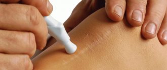
- lubricating the seam with antiseptic agents that do not contain alcohol (“Fukorcin”, “Betadine”);
- application of ointments (Levosin, Vulnuzan, Kontraktubeks and others);
- inclusion of vitamins in the diet.
If suppuration appears in the suture area, you need to treat it with antiseptic and alcohol-containing agents, for example, iodine. In addition, in these cases, antibiotics and anti-inflammatory drugs are prescribed. In order to speed up the healing of stitches, traditional medicine recommends making compresses with an alcohol tincture of larkspur. Only the roots of this herb are suitable for its preparation. They are washed well from the soil, crushed in a meat grinder, put in a jar and filled with vodka. The tincture is ready for use after 15 days. For a compress, you need to dilute it with water 1:1 so that the skin does not get burned. There are many folk remedies for healing wounds and scars after surgery. Among them are sea buckthorn oil, rosehip oil, mumiyo, beeswax, melted with olive oil. These products should be applied to gauze and applied to the scar or seam.
Complications in women whose obstetrics were performed by caesarean section are common. One of the reasons for this phenomenon is the mother’s body, weakened by pregnancy, which is unable to ensure rapid regeneration of damaged tissues.
In addition to seroma, a ligature fistula or keloid scar may occur, and in the worst case scenario, suppuration of the suture or sepsis. Seroma in women giving birth after a cesarean section is characterized by the fact that a small dense ball with exudate (lymph) inside appears on the suture. The reason for this is damaged blood vessels at the site of the incision. As a rule, it does not cause concern. Seroma of postoperative suture after cesarean does not require treatment. The only thing a woman can do at home is to treat the scar with rosehip or sea buckthorn oil to speed up its healing.
As mentioned earlier, seroma can occur after plastic surgery, but the most common are mastectomy and abdominoplasty. The formation of serous fluid occurs in almost 15% of all cases of mastectomy, and this is a fairly high chance of complications. Naturally, breast surgery leads to the most common factor in the accumulation of serous fluid, namely the spread of lymph nodes and their number in this area of the body. During breast surgery, a large dissection of the skin occurs, which affects not only a large number of blood vessels, but also lymph nodes.
As a result, already at the healing stage, due to the occurrence of an inflammatory reaction, serous fluid appears under the skin. Before performing a mastectomy, doctors warn their patients about the possibility of seroma. When undergoing abdominoplasty, the chances of fluid accumulation under the skin increase even more, because here seroma appears in almost half of the cases of plastic surgery.
In fact, the reason is identical, because when cutting the skin on the abdomen, doctors touch a large number of blood vessels and lymph nodes, which, of course, lead to further inflammatory processes.
It is always better to prevent complications from developing.
To prevent the formation of subcutaneous fluid, it is enough to follow the recommendations of surgeons:
- Immediately after the operation, a load of up to 1 kg is placed on the suture . Salt or sand can be used as cargo.
- In the first three days, traditional surgical drainage is installed.
- You should take antibacterial drugs .
- Do not perform abdominoplasty if there is a thick layer of subcutaneous fat with an index of more than 5 cm. If it is more than 5 cm, then liposuction should be performed first.
- Point effect on soft tissues . Electrocoagulation should be applied in isolation, only to bleeding vessels. Do not put pressure on soft tissues or stretch them.
- Use high-quality compression garments . This creates good compression and fixation, which prevents displacement of the skin-fat area.
- Physical rest for 3 weeks.
If recommendations and preventive measures are followed, the probability of fluid formation is 10%. Even if it appears, treatment of seroma after surgery is easy and quick.
Suppuration . Bacteria multiply very quickly in the serous fluid and the risk of suppuration is very high. Any infection - sinusitis, tonsillitis - can cause wound infection, as it spreads through the lymph and blood.
Mucosal formation . Appears during a long course of the disease, if the seroma does not go away after surgery. It is formed both on the skin-fat flap and on the abdominal wall. If the formation of a seroma is not recognized in time, an isolated cavity with fluid will appear.
This long-term condition makes the skin mobile relative to the peritoneum. Such a seroma can exist for a very long time until events occur that provoke the manifestation of this formation.
Symptoms can often include an enlarged belly. If you start this process, suppuration will begin. The only way to get rid of such a cavity is through surgery.
If the seroma is not diagnosed for a very long time and the seroma of the postoperative suture is not treated in a timely manner, this can lead to deformation of the skin-fat area and thinning of the fiber, which will affect the appearance of the skin.
The occurrence of seroma after surgery is not taken into account by many, but this can ultimately lead not only to discomfort, but also to serious illnesses or simply deformation of the skin. The removal of serous fluid is quick and painless, so this should not be put off for a long time. It is easiest to prevent the occurrence of seroma in the early stages of formation than to carry out a second operation later.
source
Seroma is a cavity with serous fluid that forms in the area of a postoperative suture or after severe injury to soft tissues and blood vessels. It is not dangerous, often resolves on its own, but often requires therapy.
The first symptom of seroma is redness of the area around the suture and swelling, and the release of yellowish fluid along the edges of the wound. Additionally, the body temperature may rise, the patient is bothered by severe pain and a feeling of fullness and heaviness.
Treatment of seroma is carried out with medications or through the installation of drainage and vacuum aspiration. When diagnosing the problem early, it is advisable to use folk remedies - aloe compresses, bran and honey cakes, cabbage leaves.
Seroma is a formation located in the tissue of the subcutaneous cavity; it looks like suppuration due to the color of the accumulated fluid: it can vary from yellow to gray and have miniature blood streaks. Most often, seroma forms in postoperative sutures, and this is always associated with the negligence of doctors and violation of sterility.
There is no separate code according to MBC 10 for this pathological condition; doctors indicate it taking into account the type of operation performed and the cause of the seroma. For example, if this happened after a cesarean section, then the medical record will indicate “0 86.0”, and this is interpreted as “suppuration of a postoperative wound and/or infiltration in its area.”
Seroma is an early postoperative complication; it usually does not persist for a long time - within 8-14 days the formation, with adequate therapy, disappears, but in some cases it can persist for one and a half to two months.
Doctors claim that the fluid accumulated in the subcutaneous cavity does not pose a threat to the patient’s health/life. But if the seroma is extensive, there is too much content, then more serious complications may develop - necrosis of surrounding tissues, sepsis, too long wound healing.
And here is more information about how to care for stitches after breast surgery.
When planning a surgical intervention, doctors take into account all the patient’s health characteristics in order to identify in advance the likelihood of developing seroma; this risk group after surgery includes patients with previously diagnosed:
- diabetes mellitus;
- hypertension;
- obesity.
This list also includes patients from older age groups whose regeneration and recovery capabilities are at a very low level. It is noted that seroma is most often diagnosed in women, because a similar complication occurs after a cesarean section, mastectomy, or abdominoplasty.
In most cases, early postoperative seroma resolves within the first 3-5 days of the rehabilitation period. As a rule, the patient is under the control of medical workers during this period, so the seroma does not develop into an advanced state. It will not resolve on its own; drug therapy or additional surgical intervention will be required.
The pathological formation in question may be small in size and not manifest itself at all for a long time, but in a certain period of time (for example, if the immune system sharply weakens, a general infectious disease develops, and so on), both inflammation and intoxication will certainly appear. If immediately after surgery seroma can be successfully treated with medication, then in advanced cases only surgery will solve the problem.
The yellow liquid is not dangerous, but only until its amount begins to increase. After all, it is the “content” of the lymphatic system, which is constantly replenished. And if no therapeutic measures are taken, the patient’s well-being will worsen, complaints of a feeling of fullness and pain in the area of seroma formation will appear. In fact, the doctor will reveal:
- skin redness;
- swelling of the site of pathological accumulation of fluid;
- increase in general body temperature to subfebrile levels.
These are signs of incipient inflammation of the skin and soft tissues surrounding the seroma. The danger is that this pathological process can rapidly progress, which leads to necrosis of the skin flap, the spread of inflammation, the introduction of an infectious agent into the cavity and sepsis.
Serous fluid usually appears for two main reasons:
- Fat deposits. During the operation, they peel off from the skin, resulting in the formation of cavities in which lymphatic fluid accumulates. It is believed that the presence of a large fat layer in a patient (more than 50 mm) is a reason for intensive weight loss before surgery or preliminary liposuction.
- The wound surface area is too large. It's simple: in this case, damage occurs to a large number of lymphatic vessels, which heal much more slowly than blood vessels. During the entire healing process, liquid oozes out of them, which accumulates in the cavities.
Seroma after surgery
But doctors also consider “individual” reasons for the formation of seroma, depending on the nature of the injury.
Post-traumatic seroma - for example, one that appears on the lower leg after an injury, is formed against the background of severe compression of tissue. It can be short-term, but at this time there is a deterioration or complete stop of lymph flow through the vessels. After medical assistance is provided, lymph rushes through the vessels with great force, so it immediately enters the tissues in large quantities.
Usually, after such injuries, seroma is immediately diagnosed in an advanced form. The reason for this is precisely the too large volume of lymphatic fluid poured into the cavity.
Seroma often occurs after breast surgery (mammoplasty, mastectomy), and the reason for this is simple - the mammary gland anatomically consists of glandular and fatty tissue, and the operation involves making large/extensive incisions that damage not only blood vessels, but also lymphatic vessels. The result is the accumulation of serous fluid under the skin already in the recovery period.
In 98% of cases, surgical interventions on the mammary gland end in the formation of seroma. Doctors warn their patients about this even at the stage of preparation for surgery.
Subcutaneous seroma is formed after electrocoagulation, a manipulation that allows blood vessels to be cauterized during surgery, which prevents the development of bleeding and most complications during the rehabilitation period. In essence, a burn occurs, in which the inflammatory process, necrosis and accumulation of fluids are inevitable.
Taking into account all the factors, doctors give a prognosis for the formation of seroma with 90% confidence. This can only be avoided by reducing the impact of electrocoagulators on tissue, but this is often impossible - the doctor works almost “blindly” and it is important for him to prevent bleeding.
There is a high probability of the formation of the pathological formation in question even after abdominoplasty, removal of a hernia, appendicitis and gallbladder bed, since during such surgical interventions the abdominal cavity is exposed to injury, in which not only a large number of lymphatic vessels are located, but also nodes of the same system.
Their damage is inevitable, and after stitching the wound and during the recovery/healing period, the lymphatic system simply does not have time to recover.
As a result, fluid from the vessels pours directly into the subcutaneous layers or soft tissues, and if rehabilitation is difficult, it accumulates in the cavities.
The second reason for the frequent diagnosis of seroma in these cases is the presence of both visceral and subcutaneous fat in the abdominal cavity.
The main symptom of postoperative seroma is the release of lymphatic fluid that occurs along the edges of the wound. It is necessarily accompanied by additional signs:
- increase in general body temperature - usually not higher than subfebrile levels, sometimes can reach 39C, which depends on the level of the patient’s immune system, the speed of the rehabilitation period, the presence of an infectious agent in the postoperative wound, and the level of progression of the inflammatory process;
- pain at the site of seroma formation - it is aching in nature, can bother you for a long time, then stop for a short period of time and reappear;
- swelling of the surgical wound site - affects the nearest skin; upon palpation, puffiness of the subcutaneous layer is also felt; it is considered one of the first signs of the formation of seroma between the lipid and subcutaneous layers of the dermis;
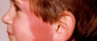
- redness of the edges of the wound, which can spread to apparently healthy skin and have a variable shade from pale pink to distinct crimson.
The patient may also be bothered by a burning pain syndrome if he changes the position of his body and a sudden rush of blood and lymph begins to the wound. The secreted liquid has no odor, but if it is present (sour, with signs of rotting), then this indicates the addition of an infection or bacterial process.
Watch this video about why seroma may occur after mammoplasty:
Seroma is a pathological process that interferes with rapid recovery after injury or surgery. Doctors must diagnose the formation, for which they prescribe:
- laboratory examination of the secreted serous fluid - it is necessary to exclude or confirm the presence of bacteria or infection in the cavity;
- laboratory testing of blood from capillaries to clarify the level of glucose, as well as the number of platelets, erythrocytes, and leukocytes;
- a blood test from a vein to exclude or confirm HIV infection, syphilis, tuberculosis;
- a smear for bacteriological examination, which is taken from the surface of the seam.
If the doctor suspects an acute inflammatory process in the soft tissues, an ultrasound examination will be prescribed.
Treatment for seroma is selected depending on the severity of the pathological process and the results of the diagnosis. Drug therapy or repeated surgery may be used. If the pathology was diagnosed at the very beginning of its development, then it is advisable to use folk remedies, but only after consultation with the attending physician.
Serous fluid is pumped out from the seroma using drainage as follows:
- The patient is given local anesthesia. These can be injections of Novocaine, Ultracaine, if there are no contraindications to the use of such medications.
- The doctor punctures the formation and expands it slightly. The puncture depth is small, and no additional damage to blood and lymphatic vessels occurs.
- A drainage is inserted directly into the open cavity - part of a rubber glove (medical), the edge of which is fixed in the skin with a plaster.
- The second end of the drainage system is placed in a special receiver, where the released serous fluid flows.
The entire system is replaced and the wound is treated with antiseptic treatment no more than once every 2-3 days. This is what makes this method of treating seroma not the most appropriate - during the period between dressings, both infectious agents and pathogenic microorganisms can penetrate into the open cavity. This will provoke expansion and worsening of the inflammatory process.
Therefore, instead of rubber drainage, modern medicine prefers to use special tubes. The receiver where the liquid flows must contain an antiseptic solution.
Most often, removal of seroma with a drainage system is used if the fluid has accumulated again, although in the recent past it was completely removed from the cavity.
Vacuum aspiration is used to remove gray as follows:
- The patient is given local anesthesia. These are injections of painkillers, if there are no contraindications to such manipulation.
- The surgeon uses an instrument to make a small incision in the area of the formed seroma.
- A tube is inserted into the resulting cavity, to which a vacuum suction apparatus is connected.
Removal of liquid continues until its color changes from yellow to ichor. The doctor immediately removes the entire system and closes the wound with sterile suture material. A sterile bandage is applied on top.
The duration of the vacuum aspiration procedure is a maximum of 30 minutes. It is advisable to perform it if the accumulation of fluid has just begun, and there are no pronounced signs of seroma yet. Healing of the suture after manipulation proceeds 2 times faster.
For treatment at home, you can use ointments with anti-inflammatory, decongestant and analgesic effects. But such therapy should still be carried out only under the supervision of a doctor - it is necessary to monitor the dynamics of the disease in order to avoid full-fledged surgical intervention in case of acute progression of the pathological process.
The following ointments can be used to treat seroma:
- Naproxen is a gel that is applied to the skin around the stitch at least 3 times a day;
- Ketoprofen - used as standard around the suture, can be applied to all swelling at least 2 times a day.
If seroma was diagnosed at an early stage of formation, and ointments began to be used immediately, then already on the 3rd day of such therapy the patient will notice a significant improvement in well-being - the pain will no longer bother you, the feeling of heaviness will disappear, the swelling of the skin will become less pronounced, and the redness will almost disappear. The usual duration of ointment therapy is 5-7 days.
Antibiotics in the treatment of seroma are used only if the pathology is already advanced, and during diagnostic measures pathogenic bacteria were identified in the serous fluid. In this case, it is advisable to prescribe:
- Erythromycin - available in tablets, belongs to the group of broad-spectrum antibiotics, taken 1 tablet 2-3 times a day;
- Cefotaxime, Ceftibuten and other drugs from the cephalosporin group are used as tablets or solutions for intramuscular administration.
Modern medicine also carries out the manipulation of introducing a solution of antibacterial drugs directly into the cavity of the seroma. This procedure is advisable if during diagnosis a high level of pathogenic microorganisms in the serous fluid was detected.
In general, antibacterial therapy lasts at least 5 days, but a significant improvement in the patient’s well-being and the condition of the postoperative suture is noted already on the 3rd day. In any case, doctors do not recommend interrupting treatment to avoid a recurrent pathological process.
Folk remedies cannot be considered full-fledged therapeutic, but after consultation with a doctor they can be used as additional therapy. If therapy is carried out exclusively by traditional medicine methods, then it is necessary to strictly monitor the state of health, well-being and appearance of the suture. In case of deterioration, immediately stop using folk remedies and seek qualified medical help.
Take 3 leaves of the plant - from the very bottom of the bush, they should be “fleshy”. They are ground in a blender or through a meat grinder into a paste and squeezed well. The remaining “cake” is placed on a bandage and applied to the postoperative wound with seroma in the form of a compress - parchment paper and polyethylene are laid on top and the pathological focus is insulated. Aloe juice is taken orally, 1 teaspoon before meals (10-15 minutes).
Treatment is carried out until complete recovery, but not longer than 7 days in a row. The duration of the compress on the wound is 2 hours. If there are no positive results, then professional drug therapy will be necessary.
They are removed from the vegetable, washed and warmed to room temperature. The thickenings on the leaves need to be cut with a knife - juice will flow out of the cuts. The leaves are applied to the wound, swollen tissues and fixed with an adhesive plaster, bandage or other bandage, but not tightly. You can lubricate the surface of the cabbage leaf with honey - this will enhance the anti-inflammatory and decongestant effects.
Prepared from bran and honey, the products are taken in proportions such that the “output” is a viscous mass. It is formed with your hands into a cake (not dense) and applied to the problem area. They don’t cover anything on top, they don’t bandage it. The time such a cake spends on the serome is 1 hour. You need to perform 2-3 procedures per day.
Already on the 2nd day of manipulation, the swelling will disappear and the redness will become less pronounced.
If a seroma has formed on a postoperative cesarean section suture, then the question of drug therapy is the last thing doctors raise. Such caution is due to the fact that the woman in labor is breastfeeding, and taking antibiotics and corticosteroids is strictly contraindicated for her.
Therefore, just after a caesarean section, it is worth resorting to folk remedies, if the seroma is not extensive and doctors do not insist on installing a drainage system or performing vacuum aspiration.
After seroma treatment, recovery is carried out in a standard manner:
- change the drainage once every 2-3 days, then completely abandon it;
- intramuscular injections of antibacterial drugs to prevent the progression of the inflammatory process;
- if necessary, take painkillers.
If the seroma is removed on time, the postoperative suture heals quickly, and the patient is discharged in the standard manner - on the 10th day after surgery.
To exclude the formation of seroma in the chest, pelvis, and abdominal cavity, doctors carry out a number of preventive measures:
- A weight is placed on the postoperative suture (immediately after the end of the surgical intervention). These could be sandbags or a heating pad with ice.
- The surgical wound is not immediately sutured, leaving a hole for installing a drainage system.
- Conducting antibacterial therapy in the early postoperative period.
- Refusal for a patient to undergo abdominoplasty if the subcutaneous fat layer is too large.
- The use of electrocoagulation is targeted, affecting only blood vessels without tension on soft tissues.
- Wearing high-quality compression garments in the early and late postoperative periods.
- Avoid physical activity for 3 weeks after surgery.
And here is more information about rehabilitation and possible complications after lipofilling.
Seroma is not considered a dangerous complication of the postoperative period, but requires monitoring by medical professionals and therapeutic measures. With proper treatment, the problem is solved within 5-7 days; in especially advanced cases, therapy can last up to 60 days and be accompanied by severe complications such as necrosis of the skin flap, sepsis, and infection of the surgical wound.
Watch this video about the causes of seroma:
source
Symptoms and diagnosis
With this complication there is almost no pain . For this reason, seroma may remain unrecognized for a long time. However, the following symptoms are observed:
- sensation of fluid transfusion in the suture area on the abdomen;
- due to edema, there may be swelling in the lower abdomen, sometimes such swelling increases and then decreases again;
- tension or tenderness in the area where lymphatic fluid accumulates;
- in a standing position, unpleasant pulling sensations may intensify;
- in the area of the largest accumulations of lymph, redness of the skin is noticeable;
- increase in body temperature to 37−37.5 and fatigue, weakness.
Seroma is diagnosed in two ways: using instruments and examination. With the latter method, the surgeon examines the patient by palpation. It immediately becomes noticeable how the liquid flows from one part to another.
The second method is ultrasound , with which the accumulated fluid is clearly visible. It collects between the fatty tissue and the anterior abdominal wall.
Treatment of serous fluid accumulation
As a rule, serous fluid after surgery resolves on its own within 4–20 days , but still, even such a minor complication cannot be left unattended. It is important to consult a doctor who will be able to advise and provide treatment at a critical moment. There are several techniques that allow you to remove serous fluid in the early stages or in case of a critical situation.
Vacuum aspiration
Vacuum aspiration is one of the most common methods for treating serous fluid. This technique, unfortunately, can only be carried out in the early stages of a complication. The essence of vacuum aspiration is to use a special apparatus to which a tube is connected and lowered to the very bottom where serous fluid has formed.
Using vacuum, the fluid is drawn out of the wound. When using this method of treatment, the old postoperative wound is not opened. In addition, pumping out serous fluid helps the skin heal faster after surgery, so many clients use vacuum aspiration just to quickly return to their normal lives.
Using drainage for seroma
Drainage is used quite often in the case of treatment for the accumulation of serous fluid. This method can be used at any stage of seroma occurrence, in contrast to vacuum aspiration. Wound secretions are pumped out using a special device, but it is important to consider the sterility of the device. That is why drains can only be used once, after which they are sent for recycling. Such drains are stored in special antiseptic solutions, and before starting work, all equipment is treated with a 0.9% sodium chloride solution .
Special devices that facilitate treatment when serous fluid occurs can be inserted through the sutures left after surgery. In addition, the device can also be removed through a small puncture, which is made near the postoperative sutures. The devices are also fixed using sutures. Doctors are required to wipe the damaged areas and nearby skin every day with a 1% solution of brilliant green. It is also necessary to constantly change the bandage.
When using a drainage tube to drain serous fluid, rubber or glass hoses to extend it. It goes without saying that even additional materials for extension must be sterile, and the vessels must be filled 1/4 full with any antiseptic solution. All this must be done in order to minimize the risk of infection through stitches or wounds. Therefore, the tubes are also replaced daily.
The serous fluid is slightly viscous, so patients are placed on their backs on a special bed so that they can, in some cases, care for the drainage tube themselves. In any case, doctors conduct constant monitoring of the patient.
The serous fluid may be quite viscous, but in this case drainage with an electric pump is used.
Treatment options
Treatment of such a complication is usually carried out in two ways - surgical and medicinal, it is more conservative. Let's consider both treatment options.
With the surgical method, a puncture is performed to remove serous fluid. In almost 90% of cases, this procedure is enough to completely remove the fluid. The surgeon performs the procedure using a syringe . The volume of pumped out liquid can vary, from 25 ml to 600 ml. It is recommended to pump out every 2-3 days to completely get rid of the problem. Usually 3-7 punctures are enough until complete elimination. If the situation is more complex, then a larger number of punctures may be needed - up to 10-15 or more. Almost always, after such manipulations, the amount of serous fluid decreases and then disappears completely.
In patients with thick fat layers, sometimes this procedure is useless and does not help solve the problem. Their seroma reaches large sizes. To do this, it is necessary to use drainage with active aspiration . With the help of drainage, the fluid constantly drains away. Gradually it becomes less and less, as a result of which the cavity closes. Everything grows together and the seroma disappears. In this case, it is also necessary to carry out drug treatment.
Preventive measures and drug treatment
Prevention is based on compliance with all the rules and principles of surgery. You should definitely wear knitted compression underwear during the postoperative period for 2 months. It can also be a bandage or an elastic bandage. In the first two weeks after surgery, the patient should refrain from any physical activity. They greatly increase the risk of developing complications.
Prevention
Timely preventive measures are always better than long-term and often painful treatment. Especially when it comes to surgical procedures. In order to prevent the development of seroma, every patient should know about the following preventive techniques:
- Upon completion of the operation, a small weight weighing up to 1 kg should be placed on the suture site. The most commonly used bags are bags of well-dried salt or regular sand.
- Installation of traditional surgical drainage for the first 2-3 days after surgery.
- Taking vitamins and minerals to increase the protective function of the immune system and speed up the process of regeneration of tissues damaged during surgery.
- Taking antibacterial drugs in the first 3 days after suturing. The type of antibiotics must be prescribed by the surgeon conducting the treatment.
You should also always remember that the seam must be made with high quality without gaps. This will ensure that there are no pockets at the junction of cut tissues and will prevent infection from entering the wound, which often becomes one of the factors in the development of seroma.