Reasons for appearance
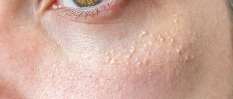
White spots under the eyes are a manifestation caused by clogging of the vellus hair follicle or sebaceous gland with keratin. It accumulates and thickens, forming whitish or yellowish lumps of a clear round shape, protruding from under the skin and hard to the touch. This problem is called milia, retention or miliary cysts or “prosyanka”.
The causes of milia are:
- lack of personal hygiene;
- use of low-quality or inappropriate care products;
- excessive use of decorative cosmetics;
- passion for chemical peeling;
- heredity;
- poor nutrition;
- burns, injuries and inflammation of the skin;
- puberty;
- oily facial skin;
- abuse of going to the solarium or exposure to the sun;
- smoking and frequent consumption of alcohol or carbonated drinks.
Barley
A stye occurs when the hair follicle of an eyelash becomes blocked. The secretion of the gland does not come out; the bacterial flora develops in the follicle sac. The edges of the eyelid swell, the point of damage to the follicle is visible. Inflammation develops around the eyes.
A stye appears on one of the eyelids. In essence, this is a purulent inflammation that requires immediate treatment. The process can quickly spread in soft tissues and affect the eye. The cause of the disease is pathogenic microflora living on the skin, as well as decreased immunity.
Possible diseases
White dots under the eyes are a rash that in itself is not a disease, but is often a sign of disorders in the body. If they occur, it is recommended to check the gastrointestinal tract, liver and gallbladder, pancreas, endocrine, cardiovascular, and autonomic nervous systems.
White nodules may indicate pregnancy, hormonal imbalance, allergic reactions, the onset of menopause, vitamin A deficiency in the body, hypo- or avitaminosis, or skin diseases (porphyria cutanea tarda, bullous pemphigoid, dystrophic form of epidermolysis bullosa, tuberculous lupus, skin sarcoidosis).
They also appear against the background of oily, thick seborrhea.
Types of white dots
Milia, or pimples under the eyes, form slowly on healthy skin. Whiteheads develop separately or in groups in the paraorbital area, on the eyelids, moving to the nose, temples, cheekbones, and cheeks. As an exception, defects are formed on the body and genitals.
Millet is easy to identify by its characteristic appearance. These are small, dense, round yellow-white nodules near the eyes. The elements range in size from half to three millimeters, rise above the skin of the face and resemble millet grains.
Do you need advice from an experienced doctor? Get a doctor's consultation online. Ask your question right now.
Ask a free question
The head of the milia does not become inflamed: it does not have a natural outlet to the surface and is not influenced by external factors. The spots near the eyes do not change size for a long time, but when bacteria enter them and the infection escalates, they transform into small pustules.
Differential diagnosis is made from the following diseases with similar symptoms:
- fibroids;
- fibrofolliculomas;
- syringomas;
- xanthelasma;
- trichodiscomas.
Milium in the eye area can be primary - genetically predetermined - and secondary, arising as a consequence of pathologies of the body. Sometimes it forms against the background of inflammation, skin injuries, or serious diseases.
Millet rarely accompanies non-malignant tumors.
Diagnostics
No special equipment, scrapings or tests are needed for diagnosis; a dermatologist-cosmetologist recognizes millet by its characteristic external signs when examining the skin. Single dots are a rare occurrence; more often they are located in clusters, covering the eyelids, temples, cheekbones and cheeks, nose, and occasionally appear on the body.
When pressed, they do not cause pain, do not itch and are not prone to inflammation if their integrity is not violated.
You need to make sure that the white lumps are milia, and not fibrofolliculomas (dome-shaped papules that do not differ in color from healthy skin or have a yellowish tint), lipomas (wen), trichodiscomas (a scattering of small whitish papules), comedones (plugs in the sebaceous glands ), syringomas (nodules formed as a result of impaired development of sweat glands).
Or xanthelasmas on the eyelids (benign tumors that are yellowish in color and soft to the touch). Only then will the treatment be correctly selected. In public clinics, consultation can be obtained free of charge; in private clinics, prices for an appointment start on average from 400 rubles.
Diagnosis of the appearance of a pimple on the eyelid under the eyelashes
Each type of neoplasm at the edge of the eye is not only an external defect, but also a symptom of some internal disease. There are the following types of acne:
- Pinguecula. It looks like a small growth that appears on the white of the apple inside the eye. It is formed due to irritation from sunlight, dirt, dust and other external influences. The function of the visual organ is not affected by the formation.
- Barley. This is an inflamed area that appears due to infection of the hair follicle. The favorite place of formation is the eyelids. Has a dense structure. The areas surrounding the stye often become red and swollen. The cause of inflammation is decreased immunity.
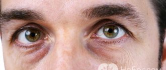
- Xanthelasma. Formation of medium density and yellow color. Develops in the area of the upper eyelid. These neoplasms can be numerous or single, and tend to increase in diameter. Factors responsible for growth are improper fat metabolism, liver disease, and high blood cholesterol levels.
- Chalazion. The growth is in the form of a nodule, dense to the touch. It is formed as a result of clogging of the excretory ducts coming from the sebaceous gland with sebum and their retention under the skin. Such pimples can appear after improper treatment of stye or from an eye infection.
- Millet and milia. These are small cysts containing keratin inside. The person does not experience pain or discomfort.
- Zhiroviki. A dense formation filled with adipose tissue. At the beginning of its development, it is often confused with a pimple, but it is impossible to squeeze it out without the help of a specialist.
To correctly diagnose pimples, the following methods are used:
- Examination by a dermatologist, surgeon and ophthalmologist.
- Assessment of the condition of blood vessels and nerve endings of the fundus.
- Measuring eye pressure.
- Checking visual function and acuity.
- Biopsy to exclude the development of a malignant process.
- Study of the condition of the eye components and tissues.
In some cases, the patient may need to consult an endocrinologist and other specialists.
Our readers recommend!
- Eye burn after eyelash extensions: signs and treatment
- What to do if the roots of your eyelashes hurt
- Demodicosis of eyelids and eyelashes in humans, demodex eye mite
When to see a doctor
White spots under the eyes are such an unpleasant phenomenon that tends to get worse if you don’t fight it. The millet does not cause any harm to health or life, but it can greatly spoil the appearance.
If the occurrence of retention cysts is caused by illness, you should not delay visiting a doctor; timely treatment will prevent further complications, and getting rid of the cosmetic defect will be cheaper. It is better to entrust treatment to a dermatologist or cosmetologist; independent removal can lead to serious consequences.
Only a dermatologist or cosmetologist can properly remove white spots under the eyes.
Rashes on the face of newborns are not uncommon; they usually go away on their own after 1-3 months and do not require intervention. In adults, cysts do not disappear without treatment, since they are not able to mature and spontaneously open.
What does a watery pimple indicate?
A transparent blister on the eyelid can appear in absolutely anyone at any time. The skin around the eyes is delicate and it is difficult not to notice this phenomenon. Know that this rash can be an alarming symptom and it is important to promptly identify the causes of its appearance.
Favorite location is the upper and lower eyelids, the edge of eyelash growth, and the inner side of the eyelid.
Why do bubbles appear on the eyes?
- Irritation. It is often provoked by hypothermia or the ingress of foreign substances and is not dangerous. No special treatment is required, other than temporarily giving up cosmetics and going out into strong winds and frost.
- Herpes. A watery growth may indicate infection with this unpleasant disease. The virus manifests itself with pain, itching, fever, and then rashes with clear and then cloudy liquid inside. Often the infection affects the inner corner and even the cornea of the eye. The patient complains of pain. Treatment is lengthy and complications can cause vision loss.
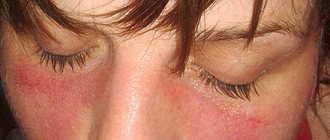
- Eczema. The preliminary symptomatic picture is rash, itching and redness. The bubble is only a harbinger of this serious and formidable disease.
There are other reasons for the appearance of bubbles on the eyes. For example, Moll cyst, papillomavirus or allergies. In each case, a complete examination and an accurate diagnosis are required.
Prevention
White spots under the eyes are a disorder of the body that is more susceptible to people with a genetic predisposition or oily skin. In these cases, the risk of cyst re-formation after removal is especially high.
However, no one is immune from this scourge, so even if there is no problem, you should follow the recommendations:
- Cleanse your face daily, choosing products based on your skin type. Scrub or exfoliate your skin 1-2 times a week. Sometimes wipe your face with a decoction of sage, chamomile, and string to refresh and disinfect.
- Reduce the consumption of salt, flour, sweet, smoked, fatty, spicy, fried foods, canned foods, carbonated drinks, avoid products with aromatic additives, dyes, and introduce fruits and vegetables into the daily menu.
- Limit exposure to the sun.
- Apply only high-quality decorative cosmetics, and be sure to wash them off before going to bed.
- Adopt a healthy lifestyle, stop smoking and do not abuse alcohol.
- If you have vitamin deficiency, do not neglect vitamin-mineral complexes. Vitamins A and E are especially important.
- Monitor your health and undergo regular examinations.
Don't miss the most popular article in the section: Face fitness for facelift, rejuvenation, muscle tone. Master class from Elena Karkukli
Treatment methods
In order not to harm yourself, you should contact a specialist with the problem. It’s not enough to get rid of white rashes; you need to follow your doctor’s instructions to avoid worsening or recurrence of the problem. Self-medication takes longer and does not always guarantee positive results.
Medications
All products are applied to cleansed skin. They help fight millet:
- Boric or salicylic alcohol (costs around 20 rubles) – they should be used to wipe each nodule point by point in the morning and evening. Healthy skin should not be touched to avoid drying it out. If an acid solution is used instead of alcohol, it should be 1% so as not to cause a burn. Noticeable improvements should appear within 10 days.
- Vishnevsky ointment (costs about 50 rubles) - squeeze a little onto a piece of cotton wool, apply to the affected area, secure with a band-aid and leave overnight. After several procedures, the “grains” should soften and can be gently squeezed out.
- Lipobase (260 rubles), Emolium (600), Retinoic ointment (300 rubles) - require spot application on the nodules themselves twice a day. Apply until the effect becomes obvious.
The duration of the course of treatment depends on the size of the rash; the more there are, the longer the recovery will take.
The advantage of folk remedies is their availability, among the disadvantages:
- Medicines are useless if milia are deep.
- The course of treatment can last several months, but does not guarantee the disappearance of all nodules.
- The products may cause irritation or an allergic reaction; in these cases, the drug should be stopped immediately.
After removing whiteheads, it is worth making masks from bodyaga; thanks to them, the wounds will heal faster and inflammation will go away. If the inflammatory process cannot be avoided, ichthyol compresses are used. Apply ichthyol ointment to a cotton pad, apply to the affected skin and secure with a band-aid. If possible, keep the compress for a day, renewing the medicine every 8-10 hours.
Improvements should be obvious after the first application.
Aspirin also has an anti-inflammatory, drying and healing effect. Crush 10 tablets, add 2-3 tbsp. warm water, stir. For better results, you can add a little liquid honey. Apply this paste to problem areas and leave for 5-10 minutes. Repeat 1, maximum 2 times a week.
https://youtu.be/u_m4PBt3ND4
Traditional methods
Medicines can be prepared with your own hands from natural raw materials. They should be used until the millet is completely eliminated. But you need to be prepared for the fact that the treatment will be long, it will take at least a month.
List of traditional methods:
- Lotion . Wash a few aloe leaves thoroughly, squeeze out the juice and mix it with 1 tbsp. alcohol Wipe the cysts with the resulting liquid before going to bed.
- Masks. When treating milia, daily use is recommended; as a preventive measure, once a week is sufficient. Viburnum: crush a handful of fresh berries with a wooden spoon, squeeze out the juice, add oatmeal, crushed with a blender, into it. Apply the paste to the white spots and leave for 45 minutes, then rinse with cool water. Cucumber. Remove the skin and seeds of one cucumber, finely chop or grate the pulp. Pour hot water, leave for at least 4 hours, wrapping the container for slow cooling. To apply the mask, you need to cut a mask from a small piece of linen or cotton with holes for the lips and eyes, moisten it in cucumber water and apply it to the face for 20-30 minutes. Paraffin. Melt the paraffin in a water bath, stirring, cool slightly, not allowing it to harden. Apply with a brush to milia and let dry, apply 2 more layers. After 20 min. remove carefully. Paraffin is contraindicated only for those who suffer from spider veins and overly sensitive skin. Milk-gelatin film mask: mix 1 tbsp. l. gelatin and milk, heat in the microwave until the gelatin dissolves, 10-15 seconds. Apply with a brush to problem areas. Wait until dry, remove. Wipe the skin with lotion. Spread half a peeled onion with honey and bake in the oven for half an hour at 100°C, crush. Apply the mixture to the affected areas in the mornings and evenings, leave for a quarter of an hour, rinse with warm water.
- Oil . Chop a small piece of propolis well and add sunflower oil. Leave for 3 days in a cool place. Treat whiteheads every morning. You can mix castor oil with tea tree oil. Treat the nodules with the mixture morning and evening.
- Scrub . Grind oatmeal with a blender, but not into flour, add 1 tsp honey to soften. baking soda and fine table salt. Apply with smooth movements along massage lines onto damp facial skin for 2 minutes. Rinse off with warm water.
- Soda peeling . Lather baby soap and apply to face. Pour baking soda onto a cotton pad. Massage your face with it for 3 minutes. Rinse off with cool water. Apply a light moisturizer.
Other methods
The most effective methods are those carried out by professionals in beauty salons.
| Way | Description of the procedure | Duration and frequency | Advantages and disadvantages | average cost |
| Electro-coagulation | Problem areas are excised with a metal loop of an electro-coagulator, heated under the influence of a constant high-frequency electric current. After the procedure, antiseptic treatment with a 5% potassium permanganate solution or chlorhexidine is required for 10 days. | Cysts can be removed in one session. This takes 10-15 minutes. | The method is famous for its simplicity, low traumatism due to point cauterization, and high efficiency. The pulses are able to penetrate into the deep layers of the skin. After the session, slight swelling and pain remain in the affected areas, which disappear after a few hours, and dense crusts appear, they disappear on their own. The main disadvantage of electro-coagulation is the likelihood of residual scars appearing. There are contraindications:
| From 70 rub. for one milium. |
| Laser | Exposure to a CO-2 (carbon dioxide) laser allows layer-by-layer clearing of rashes from any area of the face through thermal effects. Suitable for large areas affected by cysts, as well as when they are located too close to the eyes. | In one or two procedures, which last from 10 to 20 minutes, you can completely get rid of milia. | The method is painless and does not damage healthy skin. In addition to effective cleansing, it provides a bactericidal effect. After its use, there is no redness or suppuration. Laser coagulation has the highest cost among hardware methods. | From 100 rub. for each milia on the face and from 200 rubles. on centuries. |
| Mechanical cleaning | On steamed skin, the master removes millet stains using sterile instruments - a Vidal needle and a Uno spoon. | In one procedure, it is possible to remove only 10-15 milia, after which a break of several days is needed. Treatment may take a long period. | This is the most traumatic way. At the site of each removed formation, a small wound remains, which heals quickly. The advantage is the low cost of the procedure. Mechanical removal is not suitable for people with thin or overly sensitive skin and if white spots are located on the eyelids very close to the roots of the eyelashes. | From 50 rub. for one milium. |
If the milia are located close to the surface of the skin, a bath will help. The washed face should be thoroughly steamed and lightly patted with a birch or oak broom. After this, wipe your face and hands with alcohol and carefully squeeze out the blackheads. If the contents do not come out, there is no need to press harder; it is better to postpone the attempt until next time.
Don't miss the most popular article in the section: Facial massage according to the system of the Japanese doctor Asahi Zogan.
Possible complications
After eliminating a cosmetic defect, the skin needs restoration. After 4 days, it’s time to make this mask: mix 25 g of yeast with 1 tbsp. l. liquid natural honey, lemon juice and hydrogen peroxide (3%) until smooth. Keep the mask on your face for half an hour. Carry out the procedure twice a week.
If there are rashes around the eyes, you should not squeeze or pierce them at home, this is fraught with complications:
- Causing pain.
- Inflammation of wounds.
- After healing, scars will remain.
- Delicate skin is easily injured.
- Allergic reactions are possible when using traditional medicine without consulting a dermatologist.
Whiteheads are characterized by non-inflamed heads due to the fact that their contents are hidden under the skin and have no contact with the external environment. If left undisturbed, they rarely increase in size. If you puncture a miliary cyst yourself, there is a risk of introducing microorganisms into it, which will provoke inflammation with the appearance of an abscess. In this case, ointment with Erythromycin will help.
After mechanical removal, it is undesirable to use decorative cosmetics for 2-3 days.
Milia can appear at any age. White dots under the eyes are a nuisance that can affect self-esteem and cause complexes, so it is advisable to get rid of them in a timely manner. To avoid relapse, avoid stressful situations, monitor your appearance, nutrition and health.
Author: Liliya Razzhivina (Meerjungfrau)
Article design: Oleg Lozinsky
Types of skin diseases around the eyes and eyelids
Among the huge number of diseases caused by inflammation of the eyelids, several of the most common groups can be distinguished:
- bacterial lesions of the skin of the eyelids (abscess of the eyelid, phlegmon);
- inflammatory diseases of the glands and edges of the eyelids (blepharitis, barley, chalazion, meibomitis, demodicosis);
- violations of the shape and position of the eyelids (entropion, eversion, ptosis, lagophthalmos);
- allergic diseases (eczema, dermatitis, angioedema);
- developmental abnormalities of the eyelids and tumors (coloboma, papilloma, lipoma, nevus).
Bacterial lesions of the skin of the eyelids
Abscess of the century
An abscess is inflammation and redness of the eyelid with the formation of a cavity filled with pus. Often an abscess appears after an infected eyelid wound.
Causes of eyelid abscess:
- ulcerative blepharitis;
- boils;
- barley;
- purulent processes in the paranasal sinuses and orbit of the eye.
Sometimes the abscess opens on its own, and the inflammation subsides. But in most cases, a non-healing fistula appears at the site of inflammation.
Treatment of an abscess is sanitation of the eyelids and opening of a purulent formation on the eyelids with the mandatory use of antibiotics and sulfonamides.
Phlegmon
Cellulitis is an extensive inflammation of the tissues of the eyelid that occurs against the background of untreated stye. It is characterized by fever, headache, severe swelling of the eyelid and redness of the skin.
Treated with antibacterial agents, antihistamines.
Inflammatory diseases of the glands and edges of the eyelids
Blepharitis
Blepharitis is an inflammation of the eyelids, which causes redness, burning, itching and pain on the skin.
The following types of blepharitis are distinguished: allergic, meibomian, demodectic, infectious, scaly. Read more about blepharitis here.
Barley
Barley is an acute, infectious inflammation of the eyelids, resulting from pathogens entering the eyelash hair follicle or the sebaceous gland of the eyelid. Visually, stye looks like a small nodule on the eyelid.
You can read about how to properly treat stye on this page.
Meibomyitis and chalazion
Meibomyitis and chalazion are diseases associated with the appearance of a pathological process in the meibomian glands.
Meibomia appears when the duct of the sebaceous gland of the eyelid is clogged. Characterized by an acute course of the disease.
Chalazion is a chronic form of meibomitis. If a chalazion is left untreated for a long time, it turns into an eyelid cyst. Sometimes, for diagnostic purposes, it is necessary to puncture the eyelid cyst to identify the nature of the neoplasm.
Treatment and removal of a cyst on the eyelid occurs using classical techniques or laser treatment.
For more information about the symptoms and treatment of this pathology, see here.
Demodicosis
A disease of the eyelids caused by the Demodex mite. It is characterized by the appearance of yellow mucus in the eyes, gluing and breaking of eyelashes, itching and burning of the eyes.
To cleanse the eye from foci of infection, cleaning (sanitation) and rinsing of the lacrimal ducts are used. Read about the treatment of demodicosis on this page.
1 Diseases of the skin around the eyes and eyelids. Diagnosis and treatment
2 Diseases of the skin around the eyes and eyelids. Diagnosis and treatment
3 Diseases of the skin around the eyes and eyelids. Diagnosis and treatment
Violations of the shape and position of the eyelids
Turn of the century
When the eyelid is inverted, its main edge is turned onto the eyeball. Because of this, the eyelashes touch the surface of the cornea and conjunctiva, the eyes become red, irritated, and tears constantly flow from them.
Causes of pathology:
- convulsive or spastic contractions of some parts of the orbicularis oculi muscle;
- cicatricial contractions of the conjunctiva and cartilage of the eyelid that occur in certain chronic eye diseases (for example, trachoma).
The most effective method of treatment is surgery. Plastic surgery for entropion of the eyelid is also possible.
Eversion of the century
Ectropion is an inversion of the edge of the eyelid. With this inflammation of the eyelid, the conjunctiva turns out in a certain area or throughout the entire eyelid, which leads to its drying out.
The fact that the eye is constantly open, even at night, contributes to clouding of the cornea, the appearance of keratitis and other diseases.
Causes of eversion of the eyelid:
- age-related changes in the eye with sagging of the lower eyelid;
- paralysis of the orbicularis oculi muscle;
- tightening of the skin of the eyelids due to injuries, burns (cicatricial eversion), systemic diseases of the body.
First, it is necessary to eliminate the main causes that caused this disease. Surgical procedures can then be used.
Ptosis
Ptosis is an abnormally low position of the upper eyelids in relation to the eyeballs. This leads to vision defects and rapid eye fatigue. Ptosis can be congenital or acquired.
Causes of ptosis:
- damage to the oculomotor nerve;
- damage to the muscle that elevates the upper eyelid;
- strokes, encephalitis and other neurological diseases.
Treatment of the disease is mainly surgical.
Lagophthalmos
Lagophthalmos is incomplete closure of the palpebral fissure, which leads to damage and drying of the cornea and conjunctiva.
Causes of the disease:
- eyelid injuries,
- jades,
- short eyelids from birth.
In addition to treating the underlying disease, artificial tears and disinfectant drops are prescribed. In severe forms of the disease, surgical intervention with partial suturing of the palpebral fissure is used.
Allergic inflammation of the eyelids
Allergic diseases of the eyelids are accompanied by severe itching, swelling and inflammation of the eyelids.
In eczema , the skin of the eyelids is covered with papules, vesicles and pustules. Characteristic rashes appear not only on the skin of the eyelids, but also on the body. The pathology develops after suffering eyelid dermatitis or contact with allergens. At the end of the disease, crusts and serous exudate appear on the skin of the eyelids.
Characteristic signs of urticaria are swelling of the eyelids, itching and burning of the skin of the eyelids. Subsequently, blisters appear on it.
With contact dermatitis , 6 hours after contact with the allergen, swelling and inflammation of the eyelids develop with itchy papules and vesicles. Usually both eyes are affected at once.
Treatment of allergic inflammation of the eyelids is medicinal.
1 Diseases of the skin around the eyes and eyelids. Diagnosis and treatment
2 Diseases of the skin around the eyes and eyelids. Diagnosis and treatment
3 Diseases of the skin around the eyes and eyelids. Diagnosis and treatment
Anomalies of eyelid development and tumors
Coloboma
Coloboma of the eye is one of the most unpleasant diseases, which is expressed in the absence of some eye membranes. Coloboma of the eyelid is the most common. The pathology appears on the upper eyelids, but sometimes affects the lower eyelids too.
Coloboma is usually triangular in shape, where the base of the triangle is the ciliary edge of the eyelids. Since the defect affects all layers of the organ of vision, there are no eyelashes and glands in the area of coloboma.
In most cases, it is congenital, but can also occur as a result of injury and other illnesses. Another type of congenital coloboma, iris coloboma, is one of the main causes of vision loss in childhood.
Coloboma is a serious danger to the eye, as it leads to degeneration of the cornea, keratitis and other secondary diseases of the organs of vision, as well as blindness!
The most effective treatment for coloboma is surgery. Surgery for coloboma involves excision of damaged tissue and movement of a musculocutaneous flap to the site of the defect. With the help of plastic surgery, the correct edge of the eyelids is formed, which prevents the appearance of ptosis, entropion, and other complications.
Benign formations of the century
Papilloma of the eyelids
Papilloma appears on the lower eyelid usually in people over 60 years of age.
It grows slowly, looks like gray-yellow papillae, and has a spherical or cylindrical shape. In 1 case out of 100, papilloma can turn into cancer. Treatment of eyelid papilloma is surgical.
Senile wart
A senile wart is a flat and even formation of brown, yellow or gray color. It grows in older people along the lower ciliary edge of the eyelids. Treatment: cryodestruction or laser removal.
Cutaneous horn
The cutaneous horn is a dirty gray formation protruding above the eyelid. Electro- and laser excision are used for treatment.
Nevi
Nevus (or mole ) is a benign formation on the skin, from light brown to black. Nevi on the eyelid can be inherited, appear due to long exposure to the sun, disruptions in the hormonal system, or after taking certain medications. Hanging moles can appear in old age. If the formation does not bother you, there is no need to treat it.
If unpleasant symptoms appear: itching, pain, growth, blurred vision, you must consult an ophthalmologist to investigate the nature of the mole and receive the necessary treatment.
Atheroma
Eyelid atheroma ( epidermal cyst ) is a painless, dense formation that appears on the eyelid due to blockage of the sebaceous gland ducts.
Over time, the atheroma begins to increase in size and can impair vision. In this case, surgical procedures are performed. After removal of atheroma of the eyelid, there are usually no complications.

