How to identify and treat necrosis - diagnosis and treatment of the disease
By its nature, the disease in question has quite serious consequences, since the result of necrosis is the death of individual (sometimes very large) areas of tissue.
As a result, the patient’s organs and systems will not be able to function fully in the future.
Necrosis is often the cause of death: pathological cells grow very quickly, so you should respond to the first symptoms of the disease immediately.
Diagnosis of necrosis - how to determine the form and stage of the disease?
In its development, this disease goes through 3 stages:
At this stage, certain changes take place, but they are reversible.
Necrosis of the affected cells occurs.
Pathological tissues disintegrate.
To identify necrosis, which is superficial, there are no special problems: the doctor gets acquainted with the patient’s complaints, conducts blood testing, and takes a sample of fluid from the wound surface. In some cases, if gas gangrene is suspected, an x-ray of the affected area may be prescribed (to confirm the presence of gases).
For necrosis of internal organs, the diagnostic procedure is more extensive and may include:
Effective at stages 2 and 3 of the disease. At the initial stage of the disease, even in the presence of pronounced manifestations, the disease may not be detected. With sequestration, the problem of diagnosing in the later stages may be that this pathology will be combined with osteoporosis, which is endowed with similar symptoms
- Radioisotope scanning.
It is prescribed in cases where the previous diagnostic method was unsuccessful.
To carry out this procedure, the patient is administered a medication that contains a radioactive substance. A few hours later, zones of radioactivity are detected in the patient’s body.
The area affected by necrosis, due to the lack of blood circulation in it, will be presented in the image as a “cold” spot.
Used at all stages, if bone necrosis is suspected. At an early stage of the development of this pathology, the diagnostician, when performing a CT scan, should pay attention to the presence of cystic cavities filled with fluid. The presence of such formations, when previous research methods are unfruitful; The patient's complaints will help determine the diagnosis.
- Magnetic resonance imaging.
Effective at any stage of the disease, painless, safe for the patient. Using this research method, it is possible to detect even minor errors that are associated with impaired blood circulation in the tissues of internal organs.
Treatment methods for necrosis
In the treatment of any type of necrosis, several important points are taken into account:
- Type, form of necrosis.
- Stage of the disease.
- Presence/absence of concomitant ailments.
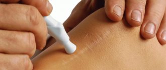
For necrosis that is localized on the skin, doctors carry out local procedures + general treatment.
If a patient is diagnosed with dry necrosis, with superficial lesions, treatment measures will include:
Procedures aimed at drying damaged tissues:
- Use of antiseptic drugs.
- Treatment of affected tissues with a solution of brilliant green/potassium permanganate.
- Use of dressings soaked in ethyl alcohol and chlorhexidine.
Procedures aimed at eliminating dead cells. During this manipulation (necrectomy), a resection of a non-functional area is performed.
The goal of general treatment of dry necrosis is to eliminate the cause that provoked the appearance of this disease. For this purpose, medications and surgical treatment can be used to restore blood circulation.
If wet necrosis with superficial lesions is detected in a patient, therapeutic measures to eliminate the pathology will include:
Local procedures.
- Treatment of the wound surface using hydrogen peroxide.
- Drainage of edema, pockets.
- The use of dressings that are impregnated with various antiseptics.
- Application of gypsum splints.
General treatment.
- Antibiotic therapy.
- The use of medications that will help prevent intoxication of the body.
- The use of drugs that help strengthen the walls of blood vessels.
Surgical procedures.
Used if measures taken to eliminate wet necrosis have not yielded results. Often, the waiting period for results in general/local treatment of wet necrosis is 2 days. If no positive changes have occurred during the specified period, an operation is performed. Any unreasonable delays can cost the patient his life.
Diagnosing necrosis in a patient, which is localized in the internal organs, involves a set of therapeutic measures:
- Taking anti-inflammatory drugs (non-steroidal).
Prescribed to relieve pain. These medications help the muscles relax, which has a positive effect on restoring blood flow. Popular drugs in this category are nimulid, piroxicam, ketoprofen, diclofenac.
- Prescription of vasodilator drugs.
Used as a method of improving blood circulation to eliminate spasm of small vessels. Restrictions on taking such medications apply to cases where there has been a stroke or myocardial infarction. The list of popular vasodilators includes: trental, theonicol.
- Medicines that promote bone tissue restoration (for sequesters).
These medications include those rich in vitamin D and calcitonins.
Prescribed in cases where there is necrosis of bone tissue. Drugs in this group help restore cartilage tissue; you need to take them for a long period. These drugs are used in the later stages of the disease.
The beneficial effect when using such leeches is achieved thanks to the enzymes that they release into the patient’s body due to suction. Through these enzymes, blood clots, which are the main cause of necrosis, are resolved and blood circulation is restored. It is not recommended to use more than 2 courses of such treatment per year.
Useful in combination with other treatment methods. Massage should not be rough, cause pain or discomfort. Improper massage can worsen the condition. This treatment procedure has some contraindications that must be taken into account.
- Laser therapy, ozokerite, mud therapy.
In combination with other therapeutic measures, they help restore normal blood circulation, reduce pain, and improve well-being. Ideal for necrosis of the hip joint.
If the patient has complaints of frequent bleeding, regular fatigue, or his medical history contains information about a recent myocardial infarction or stroke, laser therapy cannot be used.
It is effective in cases where the disease in question was caused by a pinched joint. In other cases, this type of therapy is not used as a treatment for necrosis.
In the presence of bone tissue necrosis, without this treatment procedure it is impossible to achieve full success: bone tissue necrosis provokes muscle atrophy. A set of exercises for such gymnastics must be approved by a doctor; active dynamic exercises for necrosis are unacceptable.
Source: https://www.operabelno.ru/kak-opredelit-i-vylechit-nekroz-diagnostika-i-lechenie-bolezni/
Complications after injection
Complications may occur after an injection if:
- The rules of asepsis were violated. Consequences: infiltration, abscess, sepsis, serum hepatitis, AIDS
- Wrong choice of injection site. Consequences: poorly absorbable infiltrates, damage to the periosteum (periostitis), blood vessels (necrosis, embolism), nerves (paralysis, neuritis)
- Incorrect injection technique. Consequences: needle breakage, air or drug embolism, allergic reactions, tissue necrosis, hematoma
Let's take a closer look at these types of complications.
Skin necrosis after surgery - causes, symptoms, treatment
- Causes
- Symptoms
- Postoperative necrosis
- Treatment methods
Skin necrosis is a dangerous pathology in which some tissue in the body dies.
Necrosis develops as a result of circulatory disorders, and also due to the fact that viruses and bacteria negatively affect the skin. Several types of necrosis can be defined: toxigenic, traumatic, ischemic, trophoneurotic.
It all depends on the characteristics of the structure of tissues and organs. How to properly treat the disease? Is it dangerous?
Causes
The pathology can develop subsequently from gangrene, myocardial infarction, and also due to bedsores. The skin is affected due to physical, chemical injuries, during allergies. No less dangerous are post-infectious necrosis and bedsores. They appear due to impaired blood circulation, metabolism, and failure to comply with basic hygiene rules by bedridden patients.
Necrosis can develop after an injection, when a large dose of medication is administered; subsequently, arteriolospasm first occurs, and over time, tissue hypoxia. Is it possible to prevent skin necrosis? In this case, the drug + Novocaine is administered. You can also apply cold to the injection site.
Symptoms
When skin necrosis occurs, the following unpleasant symptoms occur:
- No sensitivity.
- The skin becomes numb, pale, and sometimes even turns blue and black.
- The condition is deteriorating sharply.
- The temperature is rising.
- The pulse quickens.
- Hyperemia develops.
If unpleasant symptoms suddenly appear, and antibacterial drugs are ineffective, you should urgently consult a doctor to prevent complications of a necrotizing infection. Particularly dangerous are severe pains in the skin over the affected area; they can be the first symptom of gangrene.
Postoperative necrosis
To protect against various complications, the hospital carefully observes sterility measures, but patients who have hypertension and diabetes are most at risk. The first symptoms of the disease appear 3 days after surgery.
Along the suture, marginal necrosis develops. If the doctor sees changes, he carefully monitors the crust that covers the wound. In case of deep tissue necrosis, if the sutures begin to separate, necrectomy is performed. The edges of the wound can be cleaned with enzyme ointment or gel. After the wounds heal, a secondary suture is applied.
Skin necrosis after surgery develops due to impaired blood circulation, as well as due to significant tissue detachment and infection, which develops due to the formation of a hematoma.
Treatment methods
Pathology develops when dangerous bacteria get under the skin, resulting in skin necrosis. Bacterial gangrene is dangerous because it is caused by a non-hemolytic type of streptococcus. The streptococcal form of gangrene is caused by toxic bacilli.
If the infection progresses rapidly, severe intoxication occurs. Sometimes the skin suffers greatly after an insect bite, as well as after a minor injury, when sterility is compromised. Less commonly, necrosis is a consequence of paraproctitis or perianal abscess.
To find out about necrosis in a timely manner, a computed tomography scan is performed. The doctor plays it safe and always suggests doing a biopsy to determine histological changes.
Attention! Patients with necrosis are examined by a surgeon, resuscitator, and infectious disease specialist.
Be sure to carry out intravenous therapy using Gentamicin, Clindamycin, Penicillin. Additionally, antibacterial drugs are prescribed after microbiological examination and infusion therapy.
Bacterial gangrene develops slowly, so conservative treatment methods are used initially, then the affected skin is removed through surgery. The sooner the disease is diagnosed, the better for the patient.
In addition, the following treatment methods are required:
- Treatment of the affected tissue with a solution of potassium permanganate and brilliant green.
- Bandages are applied to the affected skin, which are pre-moistened in Chlorhexidine and Ethyl alcohol.
To cure dry necrosis, the cause is first eliminated, special medications are used, and an operation is performed during which blood circulation is restored.
If the patient has wet necrosis, a slightly different treatment is prescribed:
- Local procedures.
- Wounds are treated with Hydrogen Peroxide.
- Swelling is drained.
- Antiseptic dressings are used.
- Plaster splints are used.
Medicines are used to prevent intoxication of the body. To get rid of pain, anti-inflammatory drugs are prescribed. With the help of the medicine, the muscles relax, thus restoring blood flow. In this case, Diclofenac, Nimulid, Ketoprofen are prescribed.
To improve blood circulation, it is necessary to take vasodilators. Attention! Be extremely careful with these medications if you have previously had a heart attack or stroke.
If necrosis affects bone tissue, chondroprotectors are prescribed. With their help, you can restore cartilage tissue. Medicines must be taken at a late stage of the disease. The non-traditional method of treatment with leeches helps a lot. Due to the fact that leeches release enzymes into the body, blood circulation improves.
With necrosis, massage is indispensable. The main thing is that it is not rough, does not lead to discomfort or pain, otherwise your health will worsen. Complex therapy includes ozokerite, laser, and mud treatment. These methods do an excellent job of treating necrosis of the hip joint.
On a note! To prevent muscles from atrophying, you need to perform a special set of exercises, after consulting with your doctor.
So, necrosis occurs quite often. As a rule, it is very difficult to save a person, because everything ends in gangrene, sepsis and other unpleasant consequences. Pay close attention to your health!
Source: https://medportal.su/nekroz-kozhi-posle-operacii-prichiny-simptomy-lechenie/
Necrosis of the buttocks in a girl. There was a sharp pain like a chemical burn (18+)
Kaktus.media
at its disposal a certificate based on the results of the work of specialists from the Department of Drug Supply and Medical Equipment on the drug diclofenac sodium, after an injection of which a 12-year-old girl received necrosis of the buttock.
The document noted that a serious adverse reaction report card had to be completed and sent within 48 hours. However, it was not provided independently to DLOiMT.
It is indicated that diclofenac sodium is not recommended for use in children or patients with cardiac pathology. The literature does not exclude the rare occurrence of such an adverse reaction.
The child's mother, in her explanatory note, reports a sharp, acute pain when administering the second injection of diclofenac sodium with the immediate formation of an infiltrate and redness of the skin.
The child’s mother doubts the reliability of the administration of this drug, since the picture of complications is more typical for a chemical burn.
The explanatory note confirms the words of the Minister of Health Talantbek Batyraliev, who also spoke about the picture of a chemical burn.
From the conclusion of the debriefing protocol, it follows that when performing an intramuscular injection of diclofenac, the standards of operating procedures were not met.
From the conclusion of a freelance clinical pharmacologist of the Ministry of Health, it follows that a reaction similar to Lyell’s syndrome has developed - a severe variant of allergy (local manifestation).
“However, according to the conclusion of the vascular surgeon, allergist and explanatory procedural nurse, the following conclusion can be drawn: an instantly occurring infiltrate with acute and sharp pain without the manifestation of a general reaction of the body to the administration of the drug can only be associated with contact of the drug with an aggressive effect on soft tissues, which is not a sign of an allergic nature, since apart from local manifestations there were no signs of a general allergic response of the body to the administration of the drug,” the certificate says.
It is noted that the conclusion of the clinical pharmacologist of the Ministry of Health about Lyell's syndrome of local manifestation is also excluded, since such a term has no place in the classification of this syndrome.
“Based on the fact that the patient had previously received this drug, it follows that the injection that caused a sharp local reaction does not contain diclofenac, but another unidentified drug.
Data on the administered drug are unknown, since the ampoule and medical device were not stored after the injection, and samples were not received from the pharmacy warehouses of this organization due to lack of availability of the drug.
It is not possible to conduct a quality control analysis of this drug,” the document says.
The Department of Medicines and Medical Equipment concluded that “the injection that caused this reaction does not contain diclofenac, but another unidentified drug, the clinical course and wound healing process indicate a picture of a chemical burn
“.
Be careful, photo! Not recommended for viewing by children, weak-hearted people with unstable psyches. The photo can be viewed by clicking on the word “spoiler”.
Spoiler
Let us remind you that at the National Center for Maternal and Child Health in the department of cardiorheumatology, a 12-year-old girl was given an injection of diclofenac, after which the child began to experience necrosis of the tissue of the buttock.
The girl's mother wrote a complaint to the ministry about the quality of medical care provided to the child. On the instructions of Minister Talantbek Batyraliev, an analysis was carried out on August 18 with the participation of all responsible persons, including the leadership of the National Center for Maternal and Child Welfare (NCOMCD) and the child’s parents.
During the analysis, an underestimation of the organization of medical care for the child in the above department was confirmed after the development of complications as a result of the intramuscular administration of diclofenac. “The cause of necrosis, according to clinical pharmacologists, is a local allergic reaction to diclofenac similar to Lyell’s syndrome. They did not fill out the “yellow card,” the Ministry of Health noted.
Based on the results of the analysis, the leadership of the National Center for Children and Children was instructed to take comprehensive disciplinary measures against all those responsible.
The National Center for Maternal and Child Health did not agree with this and asked to re-conduct the investigation. They believe that the cause of necrosis was a side effect of intramuscular diclofenac.
As the hospital notes, the “yellow card” and all the necessary documents have been transferred to the Department of Medicines and Medical Equipment.
The Department investigated the case and, according to the chief physician of the National Center for Maternal and Child Health Nurgul Dzhanuzakova, came to the conclusion that the cause was an incorrectly administered injection.
Back in August, doctors received reprimands for this case, and the nurse who performed the injection quit.
Source: https://kaktus.media/doc/349216_nekroz_iagodicy_y_devochki._byla_rezkaia_bol_kak_pri_himicheskom_ojoge_18.html